. 2012 May 10;159(3):393-402.
doi: 10.1016/j.jconrel.2012.01.009. Epub 2012 Jan 20.

Liposomes loaded with paclitaxel and modified with novel triphenylphosphonium-PEG-PE conjugate possess low toxicity, target mitochondria and demonstrate enhanced antitumor effects in vitro and in vivo

Affiliations

Liposomes loaded with paclitaxel and modified with novel triphenylphosphonium-PEG-PE conjugate possess low toxicity, target mitochondria and demonstrate enhanced antitumor effects in vitro and in vivo

Swati Biswas et al. J Control Release. .

Abstract

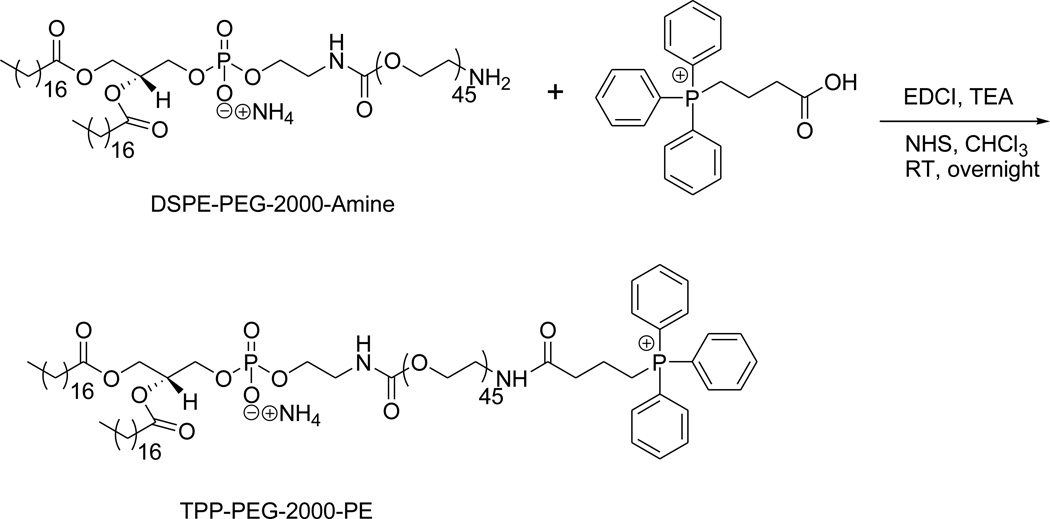
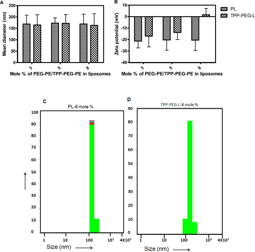
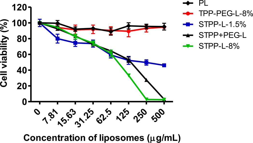
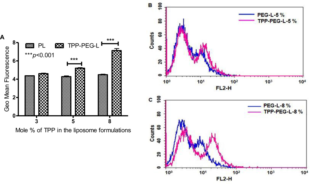
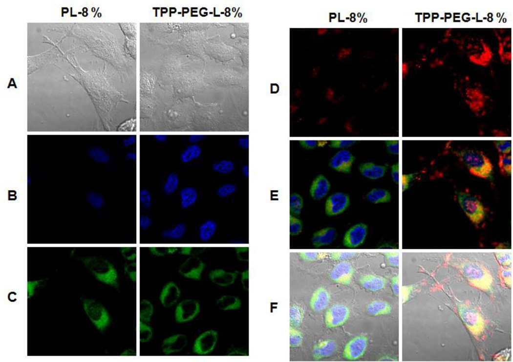
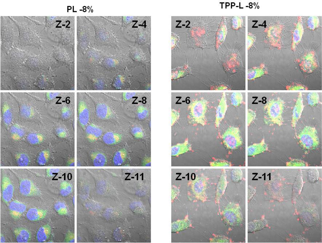
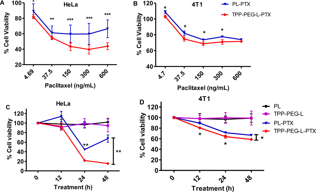
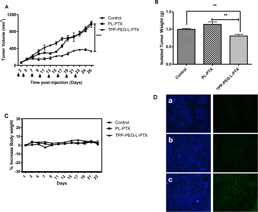
Previously, stearyl triphenylphosphonium (STPP)-modified liposomes (STPP-L) were reported to target mitochondria. To overcome a non-specific cytotoxicity of STPP-L, we synthesized a novel polyethylene glycol-phosphatidylethanolamine (PEG-PE) conjugate with the TPP group attached to the distal end of the PEG block (TPP-PEG-PE). This conjugate was incorporated into the liposomal lipid bilayer, and the modified liposomes were studied for their toxicity, mitochondrial targeting, and efficacy in delivering paclitaxel (PTX) to cancer cells in vitro and in vivo. These TPP-PEG-PE-modified liposomes (TPP-PEG-L), surface grafted with as high as 8 mol% of the conjugate, were less cytotoxic compared to STPP-L or PEGylated STPP-L. At the same time, TPP-PEG-L demonstrated efficient mitochondrial targeting in cancer cells as shown by confocal microscopy in co-localization experiments with stained mitochondria. PTX-loaded TPP-PEG-L demonstrated enhanced PTX-induced cytotoxicity and anti-tumor efficacy in cell culture and mouse experiments compared to PTX-loaded unmodified plain liposomes (PL). Thus, TPP-PEG-PE can serve as a targeting ligand to prepare non-toxic liposomes as mitochondria-targeted drug delivery systems (DDS).


Figures

Figure 1
Figure 1
Synthesis scheme for the preparation of TPP-PEG-PE.
Figure 2
Figure 2
Physico-chemical characterization of TPP-modified liposomes. A. Size of liposomes containing varied mole % of TPP-PEG-PE or PEG-PE; B. Zeta potential of TPP-PEG-L and PL; C and D. Particle size distribution of PL modified with 8 mole % PEG-PE and TPP-PEG-PE.
Figure 3
Figure 3
Survival of HeLa cells treated with TPP-modified liposomes. The cells were incubated with liposomes for 24 h, before measurement of cell-viability. TPP-PEG-L-8% were non-toxic, even at high lipid concentrations, whereas the liposomes modified with STPP induced cell death, starting at low lipid concentrations.
Figure 4
Figure 4
Quantification of the cellular uptake of TPP-PEG-L and PL modified with different mole % of TPP-PEG-PE or PEG-PE by HeLa cells by FACS analysis. HeLa cells were treated with Rh-PE-labeled TPP-PEG-L and PL for 1 h at 37 °C. A. The geometric mean of fluorescence from three separate experiments was obtained by performing statistical analysis using Cell Quest Pro software. Geometric means of the fluorescence obtained from three separate experiments were plotted. The data represents mean ± SD, n=3. (p<0.001, analyzed by the Student's t-test). B and C, histogram plots show the relative uptake levels of PL and TPP-PEG-L, presented as fluorescence intensity by FACS analysis. The PL and TPP-PEG-L were modified with 5 (B) and 8 (C) mole % of TPP-PEG-PE and PEG-PE, respectively.
Figure 5
Figure 5
Mitochondrial localization of fluorescently-labeled TPP-PEG-L compared to PL by confocal laser scanning microscopy. HeLa cells were incubated with Rh-PE-labeled TPP-PEG-L-8% and PL for 18 h and then stained with MTG and Hoechst 33342. Yellow spots in the merged pictures denote the co-localization of the liposomes within mitochondrial compartments. A. Bright field images; B. Images of nuclei stained with Hoechst; C. Mitochondria staining with MTG; D. Uptake of Rh-labeled liposomes; E. Merged picture without DIC; F. Merged picture of all (A).
Figure 6
Figure 6
Montage of z-slices obtained with confocal microscopy. The HeLa cells were incubated with Rh-labeled PL and TPP-PEG-L-8 % (red channel, laser 540 nm) for 18 h before analysis of their mitochondrial co-localization using MTG (green channel, Laser 490 nm). Hoechst 33342 was used for staining nuclei (blue channel, Laser 405 nm). Merged images of selected slices (z-2,4,6,8,10,11) from a total of 12 slices (slice thickness. 0.75 µM) are displayed.
Figure 7
Figure 7
Cytotoxicity of different preparations of PTX towards HeLa and 4T1 cells. A and B: HeLa and 4T1 cells were incubated with PL-PTX and TPP-PEG-L-PTX loaded with varied concentration of PTX (the highest PTX dose, 600 ng/mL) for 24 h. C and D: Cells were treated with PTX in different preparations at 650 ng/mL concentration for 48 h. Cell viability was measured at 12, 24 and 48 h. TPP-PEG-L-PTX resulted in significantly greater cytotoxicity than PL-PTX in both the cell lines. Empty PL and TPL used at the same lipid concentration as PL-PTX and TPP-L-PTX (50 µg/mL) were non-toxic (C and D). The data are mean ± SD, averaged from three separate experiments. *, ** and *** indicate p<0.05, p<0.01 and p<0.001 respectively, Student's t-test.
Figure 8
Figure 8
In vivo therapeutic efficacy of PTX-loaded mitochondria-targeted liposomes (TPP-PEG-L-PTX) in murine breast adenocarcinoma cell (4T1)-implanted BALB/c mice. Mice bearing 4T1-tumors received 8 i.v. injections of PTX (in PL or TPP-PEG-L-8 %) via the tail vein as indicated by the arrows. (A), Tumor growth as a function of time; saline treatment was used as control. TPP-PEG-L-PTX induced statistically significant tumor growth inhibition as compared to PL-PTX and control. (B), Postmortem tumor weights from the groups treated with TPP-PEG-L-PTX, PL-PTX or saline. (C), % increase in body weights of mice as a measure of systemic toxicity; data shown in all three graphs are expressed as mean ± SEM of n= 6 per treatment group. Overall stability in body weight was observed during the treatment period indicating no systemic toxicity of the liposomal preparations. (*, ** and *** indicate p< 0.05, 0.001 and 0.0001, Student's t-test). (D). Detection of apoptotic cells in frozen tumor sections, determined by TUNEL assay and visualized by fluorescence microscopy. The left panel shows the sections stained with Hoechst 33342 and the right panel shows the TUNEL staining. Control (PBS treatment) (a), PL-PTX (b), and TPP-PEG-L-PTX (c). Magnification- 20× objective. TPP-PEG-L-PTX induced significantly more apparent apoptosis in tumor than PL-PTX.

References

    1. Torchilin VP. Recent approaches to intracellular delivery of drugs and DNA and organelle targeting. Annu Rev Biomed Eng. 2006;8:343–375. - PubMed
    1. Shi F, Hoekstra D. Effective intracellular delivery of oligonucleotides in order to make sense of antisense. J Control Release. 2004;97:189–209. - PubMed
    1. Fulda S, Galluzzi L, Kroemer G. Targeting mitochondria for cancer therapy. Nat Rev Drug Discov. 2010;9:447–464. - PubMed
    1. Stefano JE, Hou L, Honey D, Kyazike J, Park A, Zhou Q, Pan CQ, Edmunds T. In vitro and in vivo evaluation of a non-carbohydrate targeting platform for lysosomal proteins. J Control Release. 2009;135:113–118. - PubMed
    1. Won YW, Lim KS, Kim YH. Intracellular organelle-targeted non-viral gene delivery systems. J Control Release. 2011;152:99–109. - PubMed

Publication types

MeSH terms