. 2012 Jul;11(7):844-55.
doi: 10.1128/EC.05291-11. Epub 2012 Jan 27.

Dynamic localization of Trypanosoma brucei mitochondrial DNA polymerase ID

Affiliations

Dynamic localization of Trypanosoma brucei mitochondrial DNA polymerase ID

Jeniffer Concepción-Acevedo et al. Eukaryot Cell. 2012 Jul.

Abstract

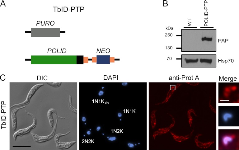
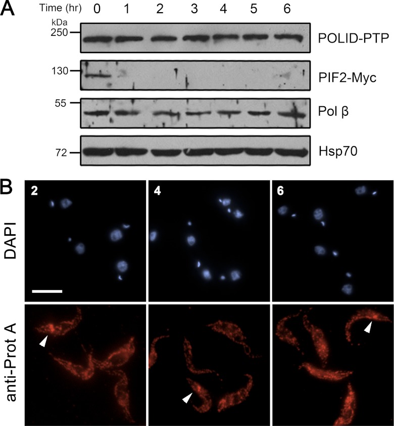
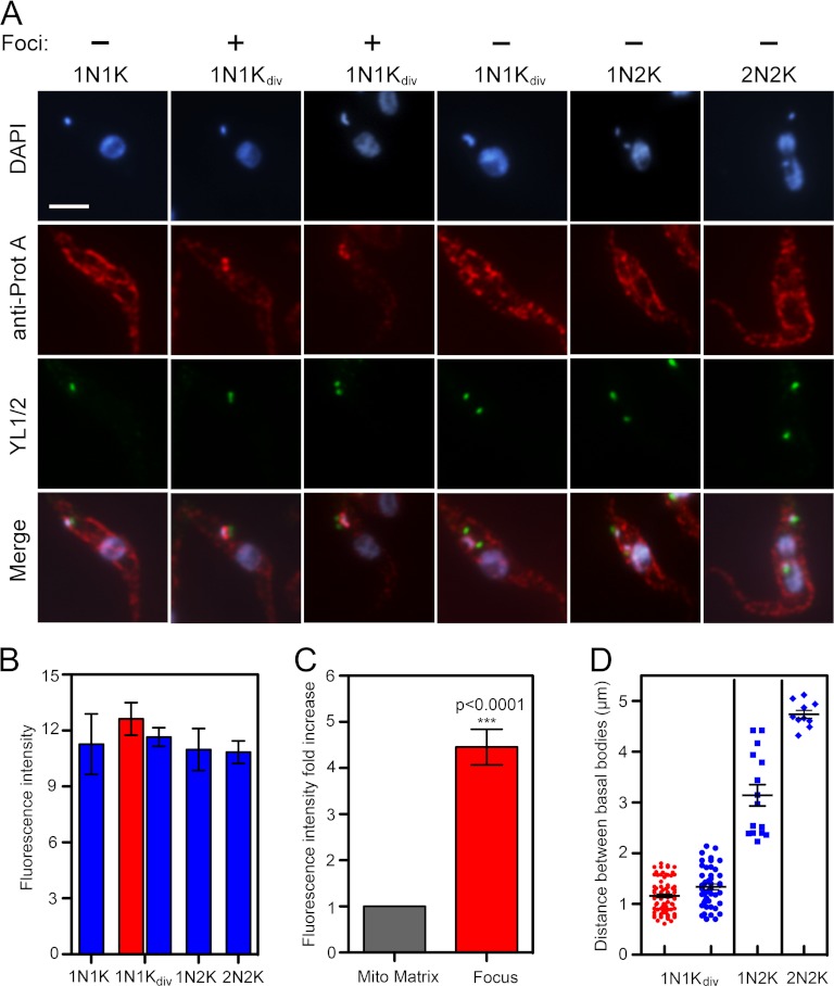
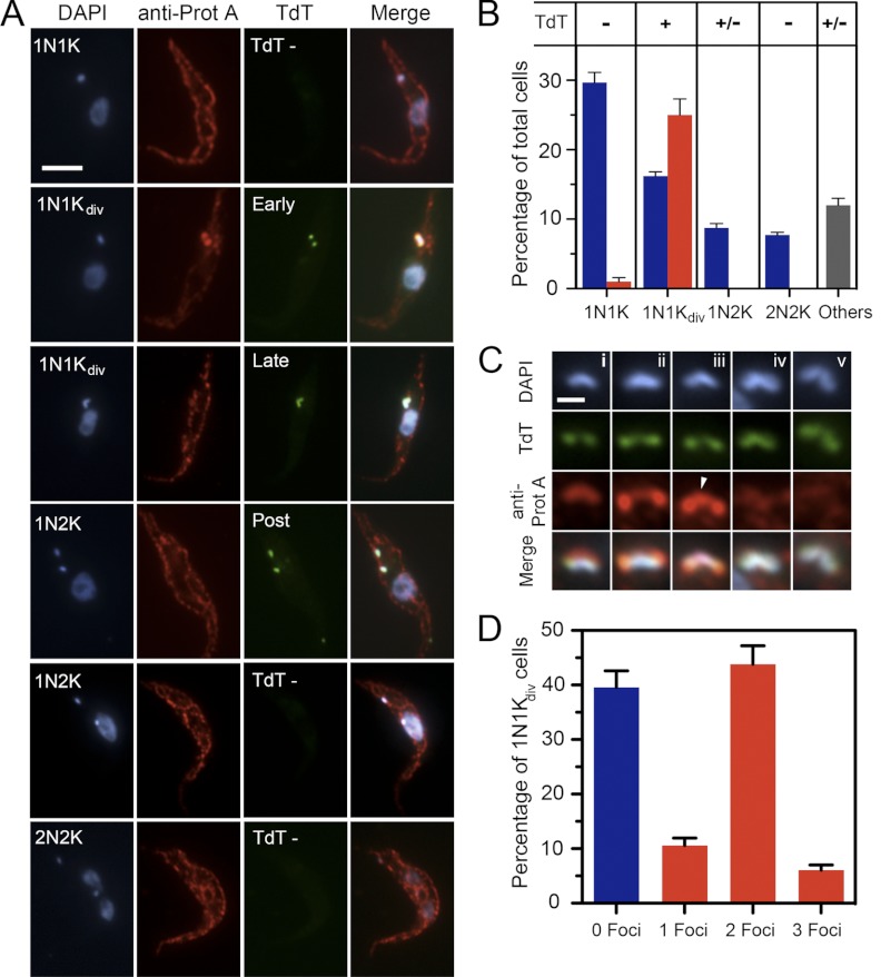
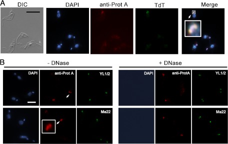
Trypanosomes contain a unique form of mitochondrial DNA called kinetoplast DNA (kDNA) that is a catenated network composed of minicircles and maxicircles. Several proteins are essential for network replication, and most of these localize to the antipodal sites or the kinetoflagellar zone. Essential components for kDNA synthesis include three mitochondrial DNA polymerases TbPOLIB, TbPOLIC, and TbPOLID). In contrast to other kDNA replication proteins, TbPOLID was previously reported to localize throughout the mitochondrial matrix. This spatial distribution suggests that TbPOLID requires redistribution to engage in kDNA replication. Here, we characterize the subcellular distribution of TbPOLID with respect to the Trypanosoma brucei cell cycle using immunofluorescence microscopy. Our analyses demonstrate that in addition to the previously reported matrix localization, TbPOLID was detected as discrete foci near the kDNA. TbPOLID foci colocalized with replicating minicircles at antipodal sites in a specific subset of the cells during stages II and III of kDNA replication. Additionally, the TbPOLID foci were stable following the inhibition of protein synthesis, detergent extraction, and DNase treatment. Taken together, these data demonstrate that TbPOLID has a dynamic localization that allows it to be spatially and temporally available to perform its role in kDNA replication.


Figures

Fig 1
Fig 1
POLID-PTP exclusive expressing cell line. (A) Diagrammatic representation (not to scale) of the TbPOLID gene locus in clonal cell line TbID-PTP. The TbPOLID coding region (green) was replaced in one allele by a puromycin resistance gene (PURO), and in the second allele the PTP sequence was fused to the 3′ end of the coding region by the targeted insertion of the pPOLID-PTP-NEO construct. Coding regions of selectable marker genes are indicated by a gray box (PURO) and blue box (NEO). The PTP sequence is indicated by a black box and introduced gene-flanking regions by small orange boxes. (B) Immunoblot analysis of whole-cell extracts of wild-type (WT) and TbID-PTP cells. A total of 5 ×10⁶ cells were loaded per lane, and tagged protein was detected with PAP reagent (top panel). The same blot was stripped and reprobed with Hsp70 antibody as a loading control. (C) Localization of POLID-PTP in an unsynchronized population. POLID-PTP was detected using anti-protein A (red), and DNA was stained with DAPI (blue). Panels in the merge column are enlargements of the area indicated by the white square. Scale bar sizes are 10 and 1 μm, respectively.
Fig 2
Fig 2
POLID-PTP protein levels and foci formation after CHX treatment. (A) Western blot detection of POLID-PTP, PIF2-Myc, Pol β, and Hsp70 protein levels following CHX treatment. Cells were harvested every hour, and 5 × 10⁶ cells were loaded into each well. (B) Immunofluorescence detection of POLID-PTP foci following CHX treatment. Cells were fixed at 2-, 4-, and 6-h intervals and stained/labeled with DAPI (blue) and anti-protein A (red). POLID-PTP foci are indicated by arrows. Scale bar, 10 μm.
Fig 3
Fig 3
Localization of POLID-PTP during the cell cycle. (A) Representative cells from an unsynchronized population. TbID-PTP cells were dually labeled with anti-protein A that detects POLID-PTP (red) and YL1/2 for the detection of bb (green). DNA was stained with DAPI (blue). Scale bar, 5 μm. (B) Mean fluorescent intensity of POLID-PTP during the cell cycle. The FI was determined in 43 cells at different stages of the cell cycle based on kDNA morphology and bb positioning. The red bar represents cells with POLID-PTP foci. (C) Fold increase in FI of POLID-PTP foci. The net FI of the mitochondrial matrix (gray bar) was calculated and normalized to determine the focus FI fold increase (red bar) (SEM, 4.4 ± 1.21; n = 10). (D) Distance between bb during the cell cycle. Measurements are from randomly selected cells (n = 149) containing 2 bb/pro-bb pairs with POLID-PTP foci (red) and without foci (blue).
Fig 4
Fig 4
POLID-PTP localization in an asynchronous cell population labeled with TdT. (A) Localization of POLID-PTP relative to the localization of replicating minicircles at the antipodal sites. Scale bar, 5 μm. (B) Distribution of POLID-PTP foci in a population of TdT-labeled cells. Cells were classified based on kDNA morphology and the presence (red bar) or absence (blue bars) of POLID foci. The 1N1Kdiv category included early and late TdT-positive cells. Others (gray bar) included cells with abnormal karyotypes, including multinucleated cells and zoids. (C) Representative images of POLID-PTP foci in 1N1Kdiv TdT-positive cells. (i) Early TdT cells with a single POLID-PTP focus. (ii) Early TdT cells with two distinct POLID-PTP foci. (iii) Early TdT cells with three foci (arrowhead). (iv and v) Late TdT cells with diffuse POLID-PTP signal. Scale bar, 1 μm. (D) The population of 1N1Kdiv cells divided into subcategories based on the number of independent foci per cell.
Fig 5
Fig 5
Detection of POLID-PTP foci in extracted cells. (A) Detergent-extracted cells prior to formaldehyde fixation labeled with TdT (green) and anti-protein A (red). Colocalization of POLID-PTP foci with replicating minicircles at the antipodal sites is represented in yellow (merge; see enlargement). Scale bar, 10 μm. (B) Detergent-extracted cells labeled with anti-protein A (red), YL1/2 (green, upper panel), or MAb 22 (green, lower panel). POLID-PTP foci and cytoskeletal components such as basal bodies and exclusion zone filaments were detected in detergent-extracted cells with (+DNase) or without (−DNase) DNase treatment. Three foci also were detected after extraction (enlargement). Scale bar, 5 μm.
Fig 6
Fig 6
Schematic representation of POLID dynamic localization throughout the T. brucei cell cycle. (A) Cells in G1 (1N1K) with a single kDNA network (blue) with POLID localized throughout the mitochondrial matrix (MM; red). These cells contained a single bb/pro-bb pair (pp/ppb; green). (B) Localization of POLID foci during kDNA duplication cycle (Sk) POLID fluorescence throughout the kDNA disk in cells with a bb/pro-bb pair close together. (C) POLID foci concentrate at the antipodal sites (antipodal sites are shown in brown). (D) A third focus accumulates at the midzone of a bilobe network. (E) Antipodal sites are not visible, and POLID is now detected in the MM. (F) KDNA daughter networks held together by the nabelschnur. POLID is only detected through the MM. (G and H) In 1N2K and 2N2K cells POLID remains dispersed throughout the MM.

Similar articles

Cited by

References

    1. Bogenhagen DF, Rousseau D, Burke S. 2008. The layered structure of human mitochondrial DNA nucleoids. J. Biol. Chem. 283:3665–3675 - PubMed
    1. Boldogh IR, et al. 2003. A protein complex containing Mdm10p, Mdm12p, and Mmm1p links mitochondrial membranes and DNA to the cytoskeleton-based segregation machinery. Mol. Biol. Cell 14:4618–4627 - PMC - PubMed
    1. Bonhivers M, Landrein N, Decossas M, Robinson DR. 2008. A monoclonal antibody marker for the exclusion-zone filaments of Trypanosoma brucei. Parasit. Vectors 1:21. - PMC - PubMed
    1. Bruhn DF, Mozeleski B, Falkin L, Klingbeil MM. 2010. Mitochondrial DNA polymerase POLIB is essential for minicircle DNA replication in African trypanosomes. Mol. Microbiol. 75:1414–1425 - PubMed
    1. Bruhn DF, Sammartino MP, Klingbeil MM. 2011. Three mitochondrial DNA polymerases are essential for kinetoplast DNA replication and survival of bloodstream form Trypanosoma brucei. Eukaryot. Cell 10:734–743 - PMC - PubMed

Publication types

MeSH terms

LinkOut - more resources