

Hematogenous extraneural metastasis of the germinomatous component of a pineal mixed germ cell tumor
- PMID: 22286191
- PMCID: PMC3493664
- DOI: 10.1007/s10014-011-0080-y
Hematogenous extraneural metastasis of the germinomatous component of a pineal mixed germ cell tumor
Abstract
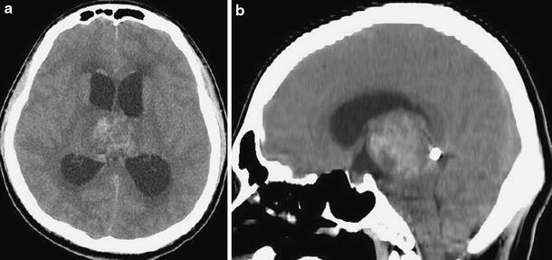
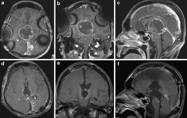
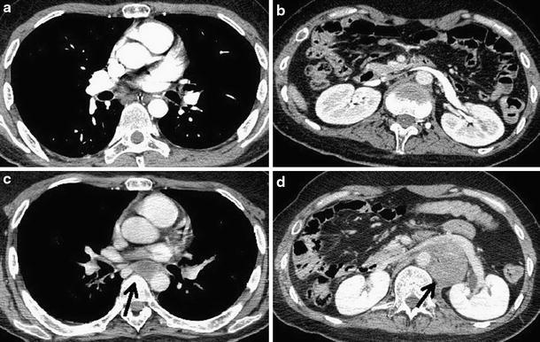
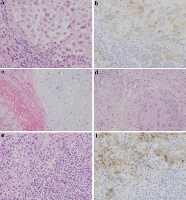
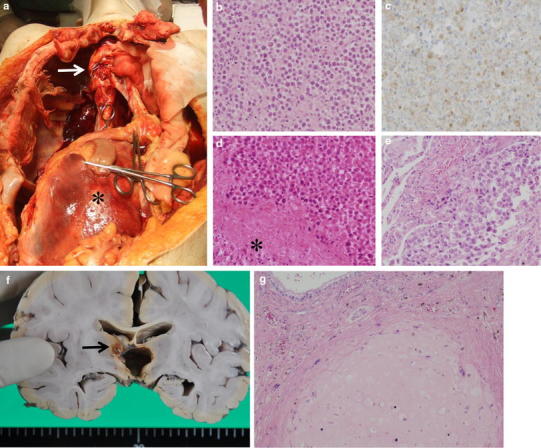
A 23-year-old man presented with a mass in the pineal region and obstructive hydrocephalus. A neuroendoscopicbiopsy for the lesion, ventriculoperitoneal (VP)shunting, and focal irradiation were conducted as initial treatment. Histological diagnosis of the biopsy specimen was germinoma. He underwent further irradiation and two tumor resections. Histological diagnosis was mature teratoma without a germinomatous component. After serial treatments, the intracranial lesion was controlled. However,14 months after presentation, extraneural lesions were confirmed in the posterior mediastinum and retroperitoneal space. The biopsy specimen of the retroperitoneal space lesion was histologically diagnosed as germinoma. Although chemotherapy with cisplatin and etoposide was undertaken,extraneural lesions ware uncontrollable and he died. At autopsy, extraneural lesions were confirmed in the posterior mediastinum, retroperitoneal space, and right lung. Histological diagnosis of extraneural lesions was germinoma.This case was considered to be a pineal mixed germ cell tumor mainly consisting of germinoma and mature teratoma,which caused hematogenous metastasis of the germinoma component. Systemic chemotherapy and irradiation for primary lesions as an initial treatment is important to cure the primary lesion and prevent extraneural metastasis.
Figures
Similar articles
-
Pure germinoma occurring 11 years after total pineal mature teratoma removal: a case report and review of the literature.Childs Nerv Syst. 2019 Dec;35(12):2423-2426. doi: 10.1007/s00381-019-04332-8. Epub 2019 Aug 5. Childs Nerv Syst. 2019. PMID: 31385089 Review.
-
Mixed germ cell tumors with abundant sarcomatous component in the temporal lobe after radiochemotherapy of neurohypophyseal germinoma: a case report.Brain Tumor Pathol. 2006 Oct;23(2):83-9. doi: 10.1007/s10014-006-0205-x. Brain Tumor Pathol. 2006. PMID: 18095124
-
Retroperitoneal teratoma with somatic malignant transformation: a papillary renal cell carcinoma in a testicular germ cell tumour metastasis following platinum-based chemotherapy.BMC Urol. 2013 Feb 12;13:9. doi: 10.1186/1471-2490-13-9. BMC Urol. 2013. PMID: 23402579 Free PMC article.
-
Endoscopic Histologic Mapping of a Mixed Germ Pineal Tumor.World Neurosurg. 2016 Nov;95:625.e1-625.e5. doi: 10.1016/j.wneu.2016.08.043. Epub 2016 Aug 23. World Neurosurg. 2016. PMID: 27554308
-
Extraneural metastasis of intracranial germinoma with syncytiotrophoblastic giant cells--case report.Neurol Med Chir (Tokyo). 1998 Sep;38(9):574-7. doi: 10.2176/nmc.38.574. Neurol Med Chir (Tokyo). 1998. PMID: 9805904 Review.
Cited by
-
Occult extracranial malignancy after complete remission of pineal mixed germ cell tumors: a rare case report and literature review.BMC Pediatr. 2023 Sep 7;23(1):447. doi: 10.1186/s12887-023-04213-9. BMC Pediatr. 2023. PMID: 37679697 Free PMC article. Review.
-
The regulatory effect of hyaluronan on human mesenchymal stem cells' fate modulates their interaction with cancer cells in vitro.Sci Rep. 2021 Oct 27;11(1):21229. doi: 10.1038/s41598-021-00754-0. Sci Rep. 2021. PMID: 34707175 Free PMC article.
-
Freiburg neuropathology case conference: a pineal region tumour in a child.Clin Neuroradiol. 2013 Dec;23(4):331-8. doi: 10.1007/s00062-013-0262-6. Clin Neuroradiol. 2013. PMID: 24141384 No abstract available.
-
Extraneural recurrence of an intracranial nongerminomatous germ cell tumor to cervical lymph nodes in a pediatric patient: Case report.Cancer Rep (Hoboken). 2022 Aug;5(8):e1586. doi: 10.1002/cnr2.1586. Epub 2021 Nov 18. Cancer Rep (Hoboken). 2022. PMID: 34796700 Free PMC article.
-
Liver metastasis in an adolescent treated for third ventricle germ cell tumor.Indian J Med Paediatr Oncol. 2014 Apr;35(2):181-3. doi: 10.4103/0971-5851.138999. Indian J Med Paediatr Oncol. 2014. PMID: 25197184 Free PMC article.
References
-
- Miyoshi Y, Omori M, Kobayashi N, Masuko T, Watanabe E, Date I. A case of pineal pure germinoma metastasized to the lumbosacral extradural space 8 years after initial therapy. Case report and review of literature. No Shinkei Geka. 2006;34:745–752. - PubMed
Publication types
MeSH terms
LinkOut - more resources
Full Text Sources
