. 2012 Jan 29;44(3):297-301.
doi: 10.1038/ng.1053.

Rare MTNR1B variants impairing melatonin receptor 1B function contribute to type 2 diabetes

Affiliations

Rare MTNR1B variants impairing melatonin receptor 1B function contribute to type 2 diabetes

Amélie Bonnefond et al. Nat Genet. .

Abstract

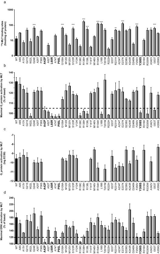
Genome-wide association studies have revealed that common noncoding variants in MTNR1B (encoding melatonin receptor 1B, also known as MT(2)) increase type 2 diabetes (T2D) risk(1,2). Although the strongest association signal was highly significant (P < 1 × 10(-20)), its contribution to T2D risk was modest (odds ratio (OR) of ∼1.10-1.15)(1-3). We performed large-scale exon resequencing in 7,632 Europeans, including 2,186 individuals with T2D, and identified 40 nonsynonymous variants, including 36 very rare variants (minor allele frequency (MAF) <0.1%), associated with T2D (OR = 3.31, 95% confidence interval (CI) = 1.78-6.18; P = 1.64 × 10(-4)). A four-tiered functional investigation of all 40 mutants revealed that 14 were non-functional and rare (MAF < 1%), and 4 were very rare with complete loss of melatonin binding and signaling capabilities. Among the very rare variants, the partial- or total-loss-of-function variants but not the neutral ones contributed to T2D (OR = 5.67, CI = 2.17-14.82; P = 4.09 × 10(-4)). Genotyping the four complete loss-of-function variants in 11,854 additional individuals revealed their association with T2D risk (8,153 individuals with T2D and 10,100 controls; OR = 3.88, CI = 1.49-10.07; P = 5.37 × 10(-3)). This study establishes a firm functional link between MTNR1B and T2D risk.


Figures

Figure 1
Figure 1. Distribution of the 40 non-synonymous MTNR1B variants identified by exon resequencing
Minor allele frequency (MAF) calculation was based on the whole sequencing dataset (7,632 individuals, including 2,186 T2D patients). Non-synonymous variants are colored as follows: blue, with MAF≥1%; green, with MAF between 0.1% and 1%; and red, with MAF<0.1%. Mutants devoid of any melatonin binding (and associated downstream signaling) are highlighted with a red flash. Mutants with impaired Gi protein-dependent signaling only are highlighted with a white flash. Mutants with impaired Gi protein-dependent signaling and ERK1/2 activation are highlighted with a black flash. Nomenclature for variants refers to functional protein sequences.
Figure 2
Figure 2. Functional characterization of wild-type and mutant MT2 receptors
(a) Expression levels in Flp-In HEK 293 cells determined by 125I-MLT saturation binding experiments. (b) Maximal Gi protein-dependent signaling. (c) EC50 values of Gi protein-dependent signaling determined in MLT dose-response curves. (d) ERK activations in the presence of 100nM MLT. The threshold line in panels (b) and (d) was defined according to the known variability of each assay. Ligand binding deficient MT2 mutants are highlighted in bold. Data are means ± standard error of at least three independent experiments, each performed in duplicate (*P<0.05; **P<0.01; ***P<0.001 significantly different from wild-type MT2). WT, wild-type; EC50, half maximal effective concentration; MLT, melatonin; 125I-MLT, 2(125I)-iodomelatonin.
Figure 3
Figure 3. Odds ratio estimates of partial or total loss-of-function versus neutral very rare variants (MAF<0.1%) for T2D risk based on MTNR1B sequencing data
For each pool of variants (purple: loss-of-function, cyan: neutral and black: altogether), we generated an estimate of the effect size and its associated standard error. The figure represents assumed normal distributions of the odds ratios (log scale) centered on the effect sizes and with standard deviations equal to the respective estimated standard error. The figure illustrates the relative impact of functional variants among all variants with MAF below 0.1%. MAF, minor allele frequency; Loss-of-function variants are variants which impair MT2 function according to our four-tier functional investigation.

References

    1. Bouatia-Naji N, et al. A variant near MTNR1B is associated with increased fasting plasma glucose levels and type 2 diabetes risk. Nat Genet. 2009;41:89–94. - PubMed
    1. Prokopenko I, et al. Variants in MTNR1B influence fasting glucose levels. Nat Genet. 2009;41:77–81. - PMC - PubMed
    1. Voight BF, et al. Twelve type 2 diabetes susceptibility loci identified through large-scale association analysis. Nat Genet. 2010;42:579–89. - PMC - PubMed
    1. Bass J, Takahashi JS. Circadian integration of metabolism and energetics. Science. 2010;330:1349–54. - PMC - PubMed
    1. Marcheva B, et al. Disruption of the clock components CLOCK and BMAL1 leads to hypoinsulinaemia and diabetes. Nature. 2010;466:627–31. - PMC - PubMed

Publication types

Substances