. 2012 Jan 29;44(3):291-6.
doi: 10.1038/ng.1076.

Five amino acids in three HLA proteins explain most of the association between MHC and seropositive rheumatoid arthritis

Affiliations

Five amino acids in three HLA proteins explain most of the association between MHC and seropositive rheumatoid arthritis

Soumya Raychaudhuri et al. Nat Genet. .

Abstract

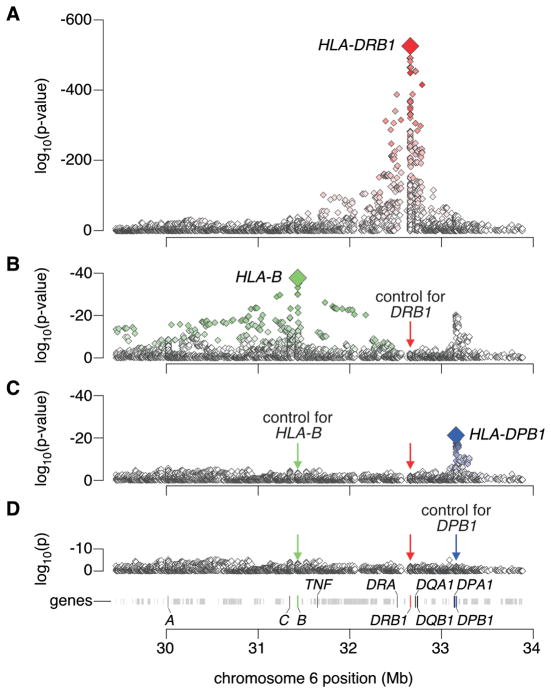
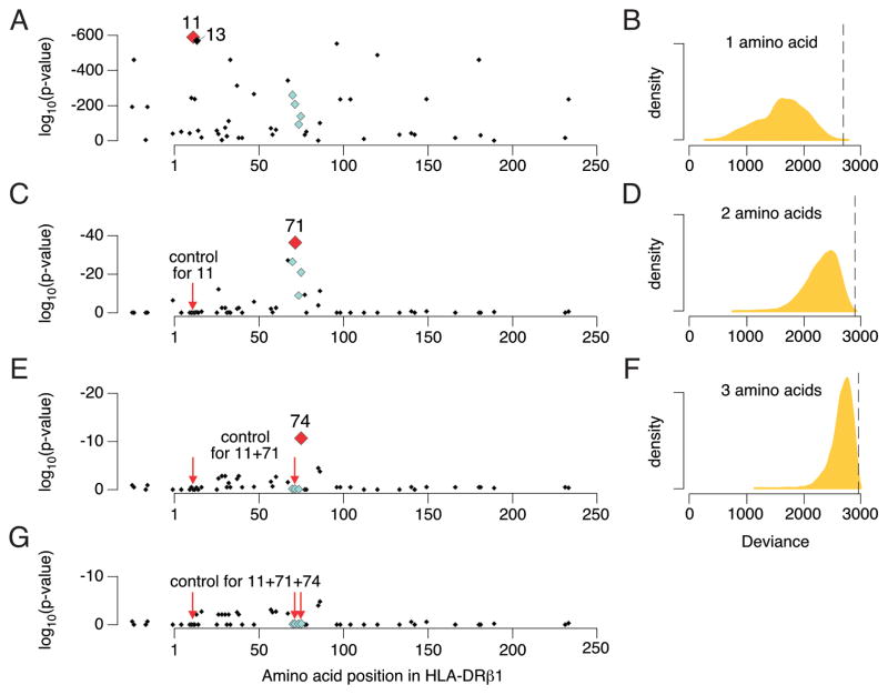
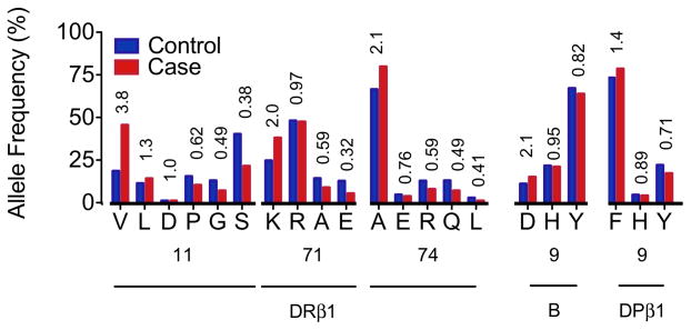
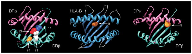
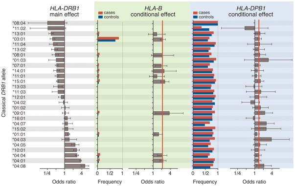
The genetic association of the major histocompatibility complex (MHC) to rheumatoid arthritis risk has commonly been attributed to alleles in HLA-DRB1. However, debate persists about the identity of the causal variants in HLA-DRB1 and the presence of independent effects elsewhere in the MHC. Using existing genome-wide SNP data in 5,018 individuals with seropositive rheumatoid arthritis (cases) and 14,974 unaffected controls, we imputed and tested classical alleles and amino acid polymorphisms in HLA-A, HLA-B, HLA-C, HLA-DPA1, HLA-DPB1, HLA-DQA1, HLA-DQB1 and HLA-DRB1, as well as 3,117 SNPs across the MHC. Conditional and haplotype analyses identified that three amino acid positions (11, 71 and 74) in HLA-DRβ1 and single-amino-acid polymorphisms in HLA-B (at position 9) and HLA-DPβ1 (at position 9), which are all located in peptide-binding grooves, almost completely explain the MHC association to rheumatoid arthritis risk. This study shows how imputation of functional variation from large reference panels can help fine map association signals in the MHC.


Figures

Figure 1
Figure 1. Association tests within the MHC to rheumatoid arthritis
(A) The major genetic determinants of rheumatoid arthritis risk map to the HLA-DRB1 gene. (B) Subsequent conditional analyses controlling for all classical HLA-DRB1 alleles reveal an independent association at HLA-B corresponding to the B*08 allele or Asp-9 in the protein. (C) Subsequent analyses that condition on HLA-DRB1 alleles and HLA-B*08 reveal an independent association for the HLA-DPβ1 Phe-9 variant. (D) Upon controlling for HLA-DRB1, HLA-B Asp-9 and HLA-DPβ1 Phe-9, no significant association signal is observed.
Figure 2
Figure 2. Association results for amino acids in HLA-DRβ1
(A) Amino acid position 11 represents the strongest association with rheumatoid arthritis (p < 10−581), followed by position 13 (p < 10−574). Shared epitope positions (70 through 74) are indicated by light-blue diamonds. (B) Distribution of deviance in 10,000 permutations of amino acid sequences across classical HLA-DRB1 alleles, where deviance is calculated as −2 times the log-likelihood for the best amino acid position. The vertical dashed line indicates the deviance for position 11 in the actual data (p = 0.0002). (C) Controlling for position 11, position 71 is significantly associated with rheumatoid arthritis (p = 5.6 × 10−38). (D) Deviance of the best two amino acid positions in 10,000 permutations. The vertical dashed line indicates the deviance for positions 11 and 71 in the actual data (p = 0.0002). (E) Controlling for positions 11 and 71, position 74 is significantly associated with rheumatoid arthritis (p = 1.5 × 10⁻¹¹). (F) Deviance of the best three amino acid positions in 10,000 permutations. The vertical dashed line indicates the deviance for positions 11, 71 and 74 in the actual data (p = 0.004). (G) After controlling for positions 11, 71 and 74, no amino acid position is significant (p > 8 × 10⁻⁴).
Figure 3
Figure 3. Effect of individual amino acids within HLA proteins
For amino acid positions 11, 71 and 74 in HLA-DRβ1, 9 in HLA-B, and 9 in HLA-DPβ1, the allele frequencies in cases (red) and controls (blue) are plotted and univariate odds ratios listed. The HLA-B and DPβ1 effects are adjusted for HLA-DRB1 alleles.
Figure 4
Figure 4. Three-dimensional ribbon models for the HLA-DR, HLA-B and HLA-DP proteins
These structures are based on Protein Data Bank entries 3pdo, 2bvp and 3lqz, respectively, with a direct view of the peptide-binding groove. Key amino acid positions identified by the association analysis are highlighted. This figure was prepared with UCSF Chimera.
Figure 5
Figure 5. Conditional haplotype analysis
Each row refers to a single classical HLA-DRB1 allele. In the left box, the main (univariate) effect is plotted as an odds ratio (with 95% confidence intervals) for each DRB1 allele (versus not having that allele), sorted in order of rheumatoid arthritis risk. In the middle box (in green), case and control allele frequencies and odds ratios are plotted for the HLA-B Asp-9 allele. In the right box (in blue), case and control allele frequencies and odds ratios (with 95% confidence internvals) are plotted for the HLA-DPβ1 Phe-9 allele. The red vertical lines indicate the aggregate effects for HLA-B and HLA-DPβ1 across all DRB1 haplotypes. The Asp-9 allele in HLA-B and the Phe-9allele in HLA-DPβ1 both have consistent effects across all HLA-DRB1 haplotype backgrounds. This suggests that these three effects are additive and independent, and not the consequence of any individual extended haplotype.

Comment in

References

    1. Isenberg D. Oxford textbook of rheumatology. Oxford University Press; Oxford; New York: 2004. p. 1278.
    1. Klareskog L, Catrina AI, Paget S. Rheumatoid arthritis. Lancet. 2009;373:659–72. - PubMed
    1. Ding B, et al. Different patterns of associations with anti-citrullinated protein antibody-positive and anti-citrullinated protein antibody-negative rheumatoid arthritis in the extended major histocompatibility complex region. Arthritis Rheum. 2009;60:30–8. - PMC - PubMed
    1. van der Woude D, et al. Quantitative heritability of anti-citrullinated protein antibody-positive and anti-citrullinated protein antibody-negative rheumatoid arthritis. Arthritis Rheum. 2009;60:916–23. - PubMed
    1. Gregersen PK, Silver J, Winchester RJ. The shared epitope hypothesis. An approach to understanding the molecular genetics of susceptibility to rheumatoid arthritis. Arthritis Rheum. 1987;30:1205–13. - PubMed

Publication types

MeSH terms