. 2012 Feb;33(2):173-81.
doi: 10.1007/s10059-012-2240-z.

The effects of rosiglitazone on osteoblastic differentiation, osteoclast formation and bone resorption

Affiliations

The effects of rosiglitazone on osteoblastic differentiation, osteoclast formation and bone resorption

Eui-Sic Cho et al. Mol Cells. 2012 Feb.

Abstract

Rosiglitazone has the potential to activate peroxisome proliferator-activated receptor-γ (PPARγ), which in turn can affect bone formation and resorption. However, the mechanisms by which rosiglitazone regulates osteoclastic orosteoblastic differentiation are not fully understood. This study examines how rosiglitazone affects osteoclast formation, bone resorption and osteoblast differentiation from mouse bone marrow. Rosiglitazone treatment not only inhibited the formation of tartrate-resistant acid phosphatase-positive cells, but also prevented pit formation by bone marrow cells in a dose- and time-dependent manner. Rosiglitazone also suppressed the receptor activator of nuclear factor (NF)-κB ligand (RANKL) receptor(RANK) expression but increased PPARγ2 expression in the cells. In addition, rosiglitazone diminished RANKL induced activation of NF-κB-DNA binding by blocking IκBαphosphorylation. Furthermore, it reduced collagen and osteocalcin levels to nearly zero and prevented mRNA expression of osteoblast-specific proteins including runtrelated transcription factor-2, osteocalcin, and type I collagen.However, mRNA levels of adipocyte-specific marker, aP2, were markedly increased in the cells co-incubated with rosiglitazone. These results suggest that PPARγ activation by rosiglitazone inhibits osteoblast differentiation with increased adipogenesis in bone marrow cells and also may prevent osteoclast formation and bone resorptionin the cells.


Figures
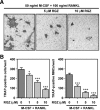
Fig. 1.
Fig. 1.
Rosiglitazone reduces the number of TRAP-positive cells in a dose-dependent manner. Bone marrow cells were exposed to the indicated concentrations of rosiglitazone in the presence of 50 ng/ml M-CSF and 100 ng/ml RANKL. The control and experimental bone marrow cells were subjected to TRAP staining (A), and the numbers of TRAP-positive cells (B) and TRAP-positive MNCs (C) were counted seven days after exposure. *P < 0.05 and ***P < 0.001 vs. the cells containing M-CSF and RANKL.
Fig. 2.
Fig. 2.
Rosiglitazone prevents the RANKL-induced pit formation in M-CSF-treated bone marrow cells. (A) Bone marrow cells were treated with 10 μM rosiglitazone in bone-coated 24-well plates and cultured for seven days in the presence of M-CSF and RANKL. Pit formation on the plate was observed under optic microscopy. (B) Bone marrow cells were also exposed to various concentrations (0–10 μM) of rosiglitazone in the presence of M-CSF and RANKL, and seven days later, the resorbed area was quantified from three independent experiments and expressed as % of the control value. ***P < 0.001 vs. the control values without rosiglitazone.
Fig. 3.
Fig. 3.
Rosiglitazone inhibits osteoclast differentiation derived from TNF-α-treated bone marrow cells and from RANKL-stimulated RAW 264.7 cells. Bone marrow cells were incubated with the indicated doses of rosiglitazone in the presence of 50 ng/ml M-CSF and 10 ng/ml TNF-α. RAW 264.7 cells were also cultured in combination with 100 ng/ml RANKL and various concentrations of rosiglitazone. After seven days of coincubation, these cells were processed for TRAP staining (A) and pit formation assays (B). The results represent the mean ± S.D. from three independent experiments. ***P < 0.001 vs. the control cells without rosiglitazone.
Fig. 4.
Fig. 4.
Rosiglitazone prevents osteoclast formation and bone resorption by bone marrow cells in a time-dependent manner. Rosiglitazone (10 μM) was added to M-CSF-treated bone marrow cultures on days 0, 1, 3, and 5 after stimulation with 100 ng/ml RANKL. Seven days after the stimulation, the cells were processed for analyses of TRAP-positive MNC formation (A) and pit formation (B). The resorbed area was also quantified from three independent experiments and expressed as % of the control value (C). **P < 0.01 and ***P < 0.001 vs. the control cells incubated with M-CSF and RANKL for seven days.
Fig. 5.
Fig. 5.
Combined treatment with rosiglitazone significantly diminishes the expression of RANK but increases PPARγ2 expression in RANKL-stimulated bone marrow cells. Bone marrow cells were incubated in α-MEM containing 50 ng/ml M-CSF, 100 ng/ml RANKL, and/or 10 μM rosiglitazone for 48 h, and then the levels of RANK (A) and PPARγ2 at mRNA (C) and protein (B) levels were determined by RT-PCR and Western blot analyses. *P < 0.05 and ***P < 0.001 vs. M-CSF treatment alone. #P < 0.05 and ###P < 0.001 vs. M-CSF and RANKL treatment.
Fig. 6.
Fig. 6.
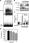
Rosiglitazone suppresses NF-κB-DNA binding and IκBα phosphorylation in RANKL-stimulated bone marrow cells. Bone marrow cells suspended in α-MEM containing 50 ng/ml M-CSF were spread onto 6-well plates, then exposed to increasing concentrations (0–10 μM) of rosiglitazone in the presence of 100 ng/ml RANKL for 48 h. Bone marrow cells were also incubated with increasing concentrations (0–50 μM) of rosiglitazone for 48 h and then processed for WST-8 assay. *P < 0.05 and ***P < 0.001 vs. the control cells without M-CSF and RANKL. #P < 0.05, ##P < 0.01 and ###P < 0.001 vs. the cultures containing both M-CSF and RANKL.
Fig. 7.
Fig. 7.
Rosiglitazone treatment decreases the contents of collagen and osteocalcin in DAG-treated bone marrow cells. Bone marrow suspension was incubated in the presence of DAG with and without the indicated concentrations (0–10 μM) of rosiglitazone, and cellular levels of collagen (A) and osteocalcin (B) were determined after ten days of incubation. *P < 0.05 and ***P < 0.001 vs. the control cells without rosiglitazone and DAG. ###P < 0.001 vs. DAG treatment only.
Fig. 8.
Fig. 8.
Combined treatment with rosiglitazone dramatically attenuates the expression of osteoblast-specific proteins but increases aP2 mRNA levels in DAG-treated bone marrow cells. Bone marrow suspension was incubated in DAG-contained medium with and without the increasing rosiglitazone concentrations (0–10 μM), and the mRNA levels of Runx2, osteocalcin, type I collagen, and aP2 were determined by real-time RT-PCR after ten days of incubation. *P < 0.05, **P < 0.01, and ***P < 0.001 vs. DAG treatment only.

Similar articles

Cited by

References

    1. Akune T., Ohba S., Kamekura S., Yamaguchi M., Chung U.I., Kubota N., Terauchi Y., Harada Y., Azuma Y., Nakamura K., et al. PPARgamma insufficiency enhances osteogenesis through osteoblast formation from bone marrow progenitors. J. Clin. Invest. 2004;113:846–855. - PMC - PubMed
    1. Ali A.A., Weinstein R.S., Stewart S.A., Parfitt A.M., Manolagas S.C., Jilka R.L. Rosiglitazone causes bone loss in mice by suppressing osteoblast differentiation and bone formation. Endocrinology. 2005;146:1226–1235. - PubMed
    1. Baek W.Y., Kim J.E. Transcriptional regulation of bone formation. Front. Biosci. (Schol Ed.) 2011;3:126–135. - PubMed
    1. Benvenuti S., Cellai I., Luciani P., Deledda C., Baglioni S., Giuliani C., Saccardi R., Mazzanti B., Dal Pozzo. S., Mannucci E., et al. Rosiglitazone stimulates adipogenesis and decreases osteoblastogenesis in human mesenchymal stem cells. J. Endocrinol. Invest. 2007;30:RC26–30. - PubMed
    1. Bradford M.M. A rapid and sensitive method for the quantitation of microgram quantities of protein utilizing the principle of protein-dye binding. Anal. Biochem. 1976;72:248–254. - PubMed

Publication types

MeSH terms