

doi: 10.1038/nmeth.1859.
In vivo protein crystallization opens new routes in structural biology
Affiliations
- PMID: 22286384
- PMCID: PMC3429599
- DOI: 10.1038/nmeth.1859
Item in Clipboard
In vivo protein crystallization opens new routes in structural biology
Nat Methods.
.
Abstract
Protein crystallization in cells has been observed several times in nature. However, owing to their small size these crystals have not yet been used for X-ray crystallographic analysis. We prepared nano-sized in vivo-grown crystals of Trypanosoma brucei enzymes and applied the emerging method of free-electron laser-based serial femtosecond crystallography to record interpretable diffraction data. This combined approach will open new opportunities in structural systems biology.
Figures
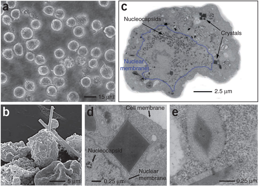
Light microscopic and EM analysis of Sf9 insect cells with embedded in vivo crystals. (a) Light micrograph of Sf9 cells infected with TbCatB virus 90 h after infection. (b) Transmission EM (TEM) micrograph of an embedded and sectioned infected Sf9 cell with crystals cut perpendicular to the long axis of the needle. Nuclear membrane is outlined in blue. (c) Scanning EM micrograph of a group of Sf9 cells infected with TbCatB virus 80 h after infection. (d) TEM micrograph of a sectioned sample, showing a crystal cut perpendicular to the long axis of the needle with surrounding membrane between nuclear and cell membrane. (e) TEM micrograph showing the lattice structure of a crystal and a longitudinal section of a second crystal (both crystals are surrounded by membrane).
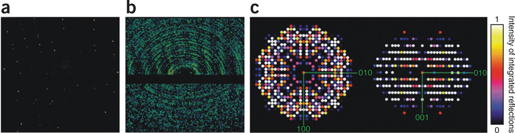
Serial femtosecond crystallography of in vivo TbCatB crystals. (a) Diffraction pattern of a TbCatB in vivo crystal recorded from a single shot of 70 fs FEL X-rays. (b) Sum of 988 single-shot FEL diffraction patterns from TbCatB crystals in different orientations. The lower panel of the detector was shifted to achieve higher resolution (Online Methods). At the edge of the detector, a maximum resolution of 7.5 Å was obtained. (c) Precession-style image of the [001] zone for TbCatB, obtained by merging SFX data from 328 in vivo crystal patterns indexed with unit cell constraints. Intensities of integrated reflections were normalized to values between 0 and 1 on a linear scale.
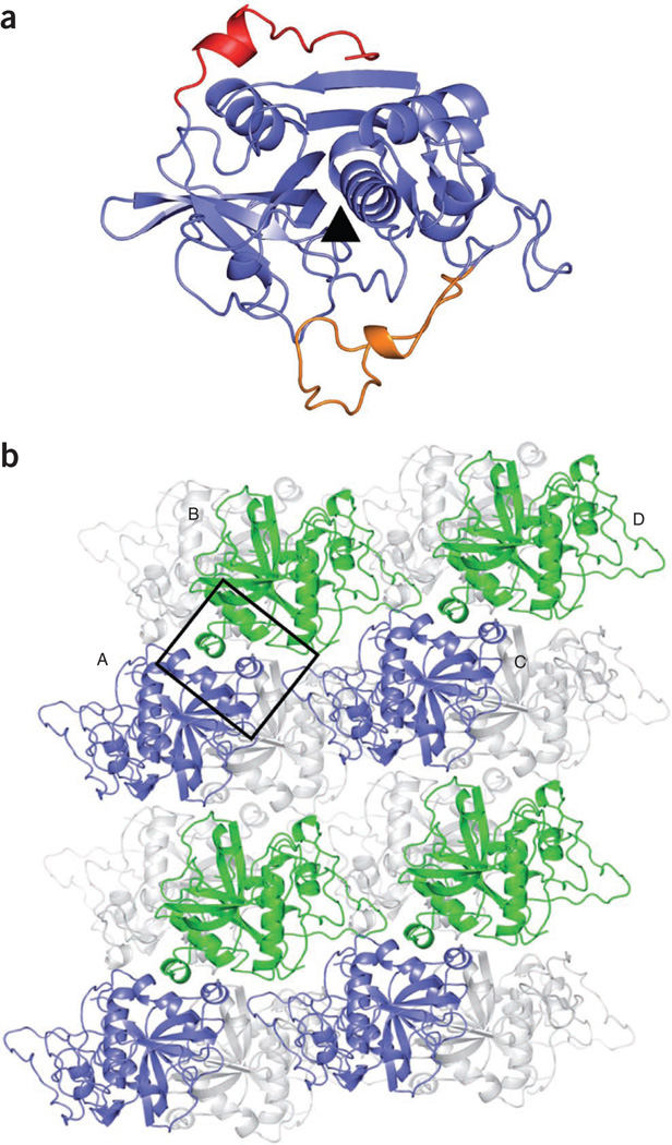
Structure of solubilized and recrystallized TbCatB solved by conventional X-ray crystallography. (a) Ribbon tracing of TbCatB, showing the pro-peptide in red and the occluding loop in orange. The black triangle indicates the position of the catalytic cleft. (b) Cartoon representation of the crystal packing. The crystals contain two molecules in their asymmetric unit and four molecules in the unit cell. This diagram of crystal contacts shows four molecules labeled A–D. In the foreground, every second TbCatB molecule is shown in blue or green, respectively. The major contact area is boxed. Gray ribbons represent additional molecules in the crystal packing.
References
Publication types
MeSH terms
Substances
Grants and funding
LinkOut - more resources
Full Text Sources
Other Literature Sources
