. 2012 Nov 22;31(47):4889-97.
doi: 10.1038/onc.2011.659. Epub 2012 Jan 30.

The PKCθ pathway participates in the aberrant accumulation of Fra-1 protein in invasive ER-negative breast cancer cells

Affiliations

The PKCθ pathway participates in the aberrant accumulation of Fra-1 protein in invasive ER-negative breast cancer cells

K Belguise et al. Oncogene. .

Abstract

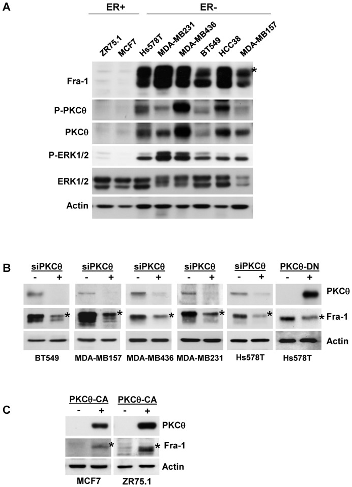
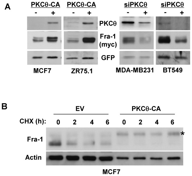
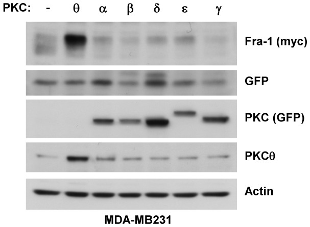
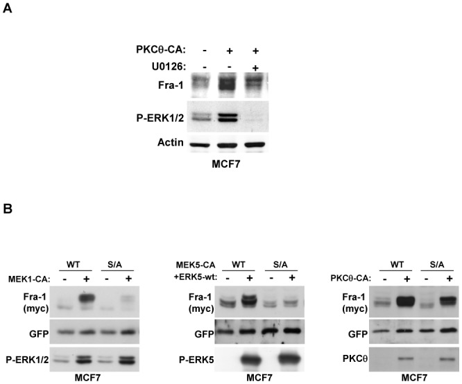
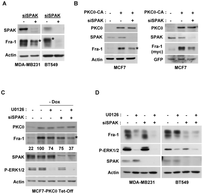
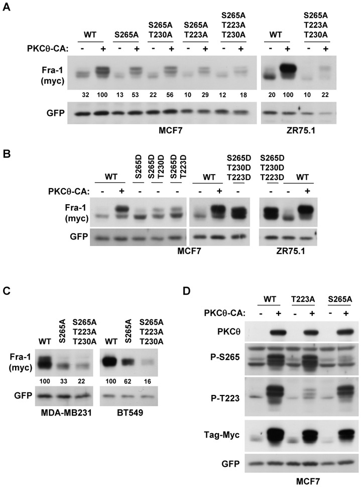
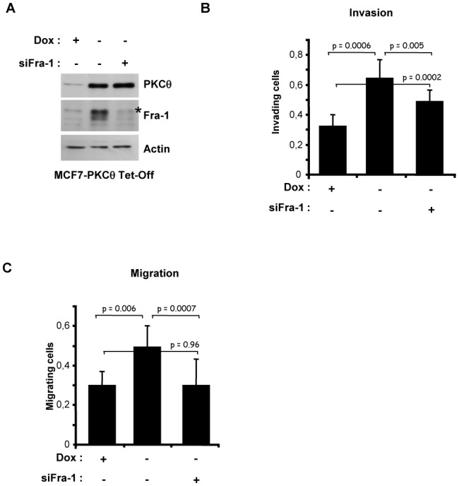
Fra-1 is aberrantly expressed in a large number of cancer cells and tissues, and emerging evidence suggests an important role for this Fos family protein in both oncogenesis and the progression or maintenance of many tumour types. Here, we show that the concentration of Fra-1 is high in invasive oestrogen receptor (ER)-negative (ER-) breast cancer cell lines, regardless of their Ras pathway status. All of the ER- cells express high levels of activated PKCθ, and the inhibition of PKCθ activity using RNA interference or the expression of a dominant-negative mutant results in a dramatic reduction in Fra-1 abundance. Conversely, the ectopic expression of constitutively active PKCθ leads to Fra-1 phosphorylation and accumulation in poorly invasive ER+ cells. This accumulation is due to the stabilisation of the Fra-1 protein through PKCθ signalling, whereas other members of the PKC family are ineffective. Both Ste20-related proline-alanine-rich kinase (SPAK) and ERK1/2, whose activities are upregulated by PKCθ, participate in PKCθ-driven Fra-1 stabilisation. Interestingly, their relative contributions appear to be different depending on the cell line studied. ERK1/2 signalling has a major role in ER- MDA-MB-231 cells, whereas Fra-1 accumulation occurs mainly through SPAK signalling in ER- BT549 cells. Fra-1 mutational analysis shows that the phosphorylation of S265, T223 and T230 is critical for PKCθ-driven Fra-1 stabilisation. Phosphorylation of the protein was confirmed using specific antisera against Fra-1 phosphorylated on T223 or S265. In addition, Fra-1 participates in PKCθ-induced cell invasion and is necessary for PKCθ-induced cell migration. In summary, we identified PKCθ signalling as an important regulator of Fra-1 accumulation in ER- breast cancer cells. Moreover, our results suggest that PKCθ could participate in progression of some breast cancers and could be a new therapeutic target.


Conflict of interest statement

Conflict of Interest

The authors declare no conflict of interest.

Figures

Figure 1
Figure 1. High PKCθ activity in ER-negative breast cancer cell lines increases Fra-1 expression
A. Whole-cell extracts (WCEs, 90 μg) from the indicated breast cancer cell lines were analysed using immunoblotting with antibodies against Fra-1, ERK1/2, phospho-ERK1/2 (T202/Y204), PKCθ, phospho-PKCθ (T538) and actin, which was used to confirm equal loading. B. The indicated ER− breast cancer cell lines were transfected with two different siRNAs (3.6 nM each) targeting the PKCθ mRNA. After two successive 48h-transfections, WCEs (90 μg) were subjected to immunoblotting for Fra-1, PKCθ, and actin, the latter being used to confirm equal loading. Alternatively, Hs578T cells were transiently transfected with 2 μg of PKCθ-DN expression vector (+) or empty vector (−) and, 48 h after transfection, WCEs (90 μg) were analysed using immunoblotting. C. The indicated ER+ breast cancer cell lines were transiently transfected with 2 μg of PKCθ-CA expression vector (+) or empty vector (−) and analysed as in B. In A, B and C, phosphorylated Fra-1 forms are indicated by an asterisk.
Figure 2
Figure 2. PKCθ increases Fra-1 protein stability in breast cancer cells
A. The indicated breast cancer cell lines were transiently transfected with 50 ng of pIRES2-EGFP vector expressing Fra-1 in the presence (+) or absence (−) of either the PKCθ-CA expression vector or siRNAs targeting the PKCθ gene as in Figure 1C and 1B, respectively. Then, WCEs (90 μg) were analysed by immunoblotting for the expression of PKCθ, Myc (detecting exogenous Fra-1) and GFP, which served as an internal standard for the normalisation of Myc-tagged Fra-1 expression. B. MCF7 cells were transiently transfected with 50 ng of pCI-Fra-1 expression vector in the presence (PKCθ-CA) or absence (EV) of the PKCθ-CA expression vector. Two days after transfection, cells were treated with 50 μg/ml of cycloheximide (CHX) for the indicated times. WCEs were then analysed using immunoblotting.
Figure 3
Figure 3. PKCθ stabilises Fra-1 protein, while other PKC subfamily members do not
MDA-MB231 cells were transfected with 50 ng of the pIRES2-EGFP vector expressing Fra-1 together with 2 μg of vector expressing the indicated wild-type PKC subfamily members or empty vector (−). Two days later, WCEs were analysed using immunoblotting for the expression of PKCθ and GFP (detecting the ectopic expression of PKCα, α:, γ, δ, and ε on the top part of the blot), Myc (detecting exogenous Fra-1), GFP (detected at the bottom part of the blot and serving as an internal standard for the normalisation of Myc-tagged Fra-1 expression), and actin.
Figure 4
Figure 4. The ERK1/2 pathway is partly involved in the stabilisation of Fra-1 protein through PKCθ signalling
A. MCF7 cells were transfected with 2 μg of PKCθ-CA expression vector (+) or empty vector (−). One day later, cells were treated with 5 μM of the pharmacological inhibitor U0126 (+) or DMSO (−) for 16 h. WCEs were then analysed using immunoblotting for endogenous levels of activated ERK1/2, Fra-1, and actin. B. MCF7 cells were transiently transfected with 50 ng of the pIRES2-EGFP vector expressing either wild-type Fra-1 (WT) or mutated Fra-1 in which serines 252 and 265 were replaced by alanine (S/A). To activate the indicated pathways, cells were co-transfected with either 2 μg of the MEK1-CA expression vector, or 1 μg of MEK5-CA plus 1 μg of the ERK5-wt expression vector, or 2 μg of the PKCθ-CA expression vector, or their corresponding empty vector (−). Two days later, WCEs were analysed using immunoblotting with antibodies against Fra-1, GFP, phospho-ERK1/2 (T202/Y204), phospho-ERK5 (T218/Y220) and PKCθ as indicated.
Figure 5
Figure 5. PKCθ stabilises Fra-1 protein through the cooperative activation of the SPAK and ERK1/2 pathways
A. MDA-MB231 and BT549 cells were transfected with 3.6 nM of siRNA targeting the SPAK gene, and after two successive 48h-transfections, WCEs (90 μg) were subjected to immunoblotting for Fra-1, SPAK and actin. B. MCF7 cells were transfected with 3.6 nM of siRNA targeting the SPAK gene, and 24 h after two successive siRNA transfections, cells were transfected with 2 μg of the PKCθ-CA expression vector (+) or empty vector (−) in the absence (left panel) or presence of 50 ng of pIRES2-EGFP expressing wild-type Fra-1 (right panel). Two days after plasmid transfection, WCEs were analysed using immunoblotting. C. Stable MCF7 cells overexpressing PKCθ-CA under tetracycline control (MCF7-PKCθ Tet-Off) were transfected with 3.6 nM of siRNA targeting the SPAK gene in the presence (+) or absence (−) of doxycycline (Dox). One day after two successive siRNA transfections, cells were treated with 5 μM of the pharmacological inhibitor U0126 (+) or DMSO (−) for 16 h. Then, WCEs were analysed using immunoblotting. The blots were scanned, and the densitometry values of Fra-1 were normalised to actin. The values relative to the non-treated cells in the absence of Dox (set at 100%) are given below each lane. D. MDA-MB231 and BT549 cells were transfected with 3.6 nM of siRNA targeting the SPAK gene. One day after two successive siRNA transfections, cells were treated with 5 μM of the pharmacological inhibitor U0126 (+) or DMSO (−) for 16 h. Then, WCEs were analysed using immunoblotting. In A, B and C, phosphorylated Fra-1 forms are indicated by an asterisk.
Figure 6
Figure 6. Serine 265 and threonines 223 and 230 are the major phosphorylation sites responsible for the stabilisation of Fra-1 by the PKCθ pathway
A. MCF7 and ZR75.1 cells were transiently transfected with 50 ng of pIRES2-EGFP vector expressing either wild-type Fra-1 (WT) or mutated Fra-1 in which the following amino acids were replaced by alanine as indicated: serine 265 (S265A), threonine 223 (T223A) or threonine 230 (T230A). To activate the PKCθ pathway, cells were co-transfected with either 2 μg of the PKCθ-CA expression vector (+) or the empty vector (−). Two days later, WCEs were analysed using immunoblotting as in Figure 2A. The blots were scanned, and the densitometry values of Fra-1 were normalised to GFP. The values relative to the cells transfected with the WT Fra-1 expression vector and PKCθ-CA (set at 100%) are given below each lane. B. MCF7 and ZR75.1 cells were transiently transfected with 50 ng of pIRES2-EGFP vector expressing either wild-type Fra-1 (WT) or mutated Fra-1 in which the following amino acids were replaced by aspartic acid as indicated: serine 265 (S265D), threonine 223 (T223D) or threonine 230 (T230D). To activate the PKCθ pathway, cells were co-transfected with 2 μg of either the PKCθ-CA expression vector (+) or the empty vector (−). Two days later, WCEs were analysed using immunoblotting as in Figure 2A. C. MDA-MB231 and BT549 cells were transiently transfected with 50 ng of pIRES2-EGFP vector expressing either wild-type Fra-1 (WT) or mutated Fra-1 in which either serine 265 or all three amino acids (serine 265 and threonines 223 and 230) were replaced by alanine. Two days later, WCEs were analysed using immunoblotting as in Figure 2A. The blots were scanned and the densitometry values of Fra-1 were normalised to GFP. The values relative to the cells transfected with the WT Fra-1 expression vector (set at 100%) are given below each lane. D. MCF7 cells were transiently transfected with 50 ng of pIRES2-EGFP vector expressing either wild-type Fra-1 (WT) or mutated Fra-1 in which either serine 265 (S265A) or threonine 223 (T223A) were replaced by alanine. To activate the PKCθ pathway, cells were co-transfected with 2 μg of either the PKCθ-CA expression vector (+) or the empty vector (−). Two days later, WCEs were analysed using immunoblotting with antibodies detecting Fra-1 only when phosphorylated at serine 265 (P265) or threonine 223 (P-223) and antibodies against PKCθ, Myc and GFP, as in Figure 2A.
Figure 7
Figure 7. Fra-1 is involved in breast cancer cell migration and invasion induced by the PKCθ pathway
MCF7 cells stably overexpressing PKCθ-CA under tetracycline control (MCF7-PKCθ Tet-Off) were transfected with 3.6 nM of siRNA targeting the FOSL1 gene in the presence (+) or absence (−) of doxycycline (Dox). Three days later, cells were either plated in 12-well plates for immunoblotting analysis after 48 h of growth (A) or subjected, in triplicate, to an invasion (B) or migration (C) assay for 48 h. Cells that migrated to the lower side of the filter were quantified using spectrometric determination of the optical density at 540 nm and normalized by the OD540 obtained with the control cells as described in Materials and Methods. The values represent the mean (± SD) of three independent experiments performed in triplicate. The p-values were calculated using the paired Student’s t-test (two-tailed). In A, phosphorylated Fra-1 forms are indicated by an asterisk.

References

    1. Shaulian E, Karin M. AP-1 in cell proliferation and survival. Oncogene. 2001;20:2390–2400. - PubMed
    1. Eferl R, Wagner EF. AP-1: a double-edged sword in tumorigenesis. Nat Rev Cancer. 2003;3:859–868. - PubMed
    1. Young MR, Colburn NH. Fra-1 a target for cancer prevention or intervention. Gene. 2006;379:1–11. - PubMed
    1. Verde P, Casalino L, Talotta F, Yaniv M, Weitzman JB. Deciphering AP-1 function in tumorigenesis: fra-ternizing on target promoters. Cell Cycle. 2007;6:2633–2639. - PubMed
    1. Mechta F, Lallemand D, Pfarr CM, Yaniv M. Transformation by ras modifies AP1 composition and activity. Oncogene. 1997;14:837–847. - PubMed

Publication types