Review
. 2012 Mar;33(3):312-23.
doi: 10.1038/aps.2011.186. Epub 2012 Jan 30.

Structure and ligand recognition of class C GPCRs

Affiliations
Review

Structure and ligand recognition of class C GPCRs

Lei Chun et al. Acta Pharmacol Sin. 2012 Mar.

Abstract

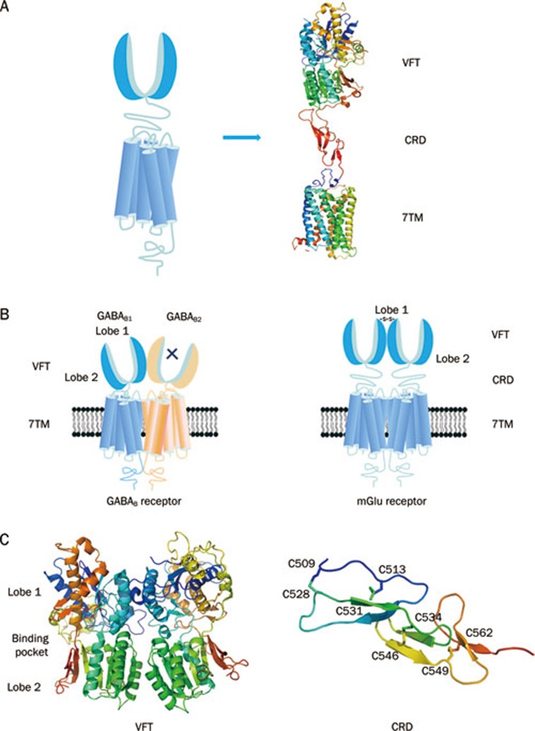
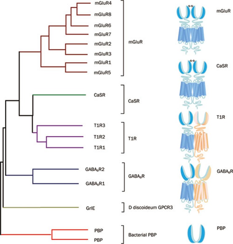
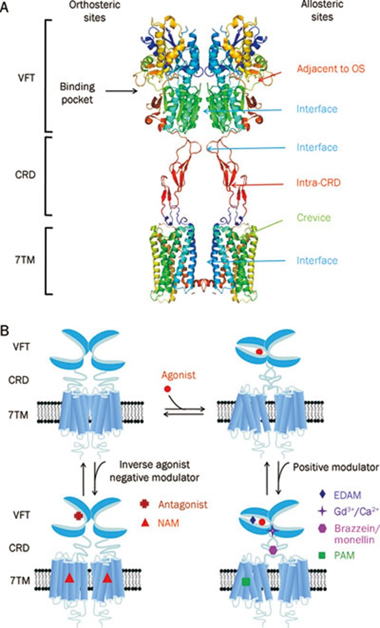
The G-protein-coupled receptors (GPCRs) are one of the largest super families of cell-surface receptors and play crucial roles in virtually every organ system. One particular family of GPCRs, the class C GPCRs, is distinguished by a characteristically large extracellular domain and constitutive dimerization. The structure and activation mechanism of this family result in potentially unique ligand recognition sites, thereby offering a variety of possibilities by which receptor activity might be modulated using novel compounds. In the present article, we aim to provide an overview of the exact sites and structural features involved in ligand recognition of the class C GPCRs. Furthermore, we demonstrate the precise steps that occur during the receptor activation process, which underlie the possibilities by which receptor function may be altered by different approaches. Finally, we use four typical family members to illustrate orthosteric and allosteric sites with representative ligands and their corresponding therapeutic potential.


Figures

Figure 1
Figure 1
Schematic structure of class C GPCRs. (A) Structural organization of class C GPCRs. Class C GPCRs have a common structure consisting of a VFT with two lobes (lobe 1 and lobe 2) separating by a cleft as orthosteric site, a 7TM and a CRD for all but GABAB receptor. The crystal structure of mGlu3 receptor (PDB ID 2E4W) was used for the VFT and CRD. The bovine rhodopsin crystal structure (PDB ID 1GZM) was used for the 7TM. (B) Schematic representation of two prototypical class C GPCRs as heterodimer (GABAB receptor), or homodimer (mGlu receptor).For GABAB receptor, the VFT is directly linked to the 7TM. Two subunits, GABAB1 and GABAB2, form an obligatory heterodimer. GABAB1 is responsible for endogenous ligands binding, while GABAB2 is responsible for G protein activating. For mGlu receptors, the VFT connects to the 7TM via CRD. mGlu receptors form homodimers which could offer two orthosteric sites per dimer. (C) The determined cystal structure for the VFT and CRD. The first solved structure is the VFT of mGlu1 receptor (PDB ID 1EWK), which shows that the VFT oscillates between an open and a closed conformation. The crystal structure of whole extracellular domain including the VFT and CRD (PDB ID 2E4W) has been solved firstly in mGlu3 receptor.
Figure 2
Figure 2
The bootstrap tree of the prototypical members from human class C GPCRs. The sequence of the VFT were aligned using the default parameters and the homologous bacterial PBPs were used as an outgroup to root the trees. Class C GPCRs form obligatory dimers. Homodimers (mGluR and CaSR) linked by a disulfide bond between their VFTs, while heterodimers (GABABR and T1R) are not covalently linked.
Figure 3
Figure 3
Great variety of ligands to modulate class C GPCRs function. (A) Schematic model of orthosteric sites and allosteric sites in mGlu-like receptor. There are four groups of allosteric sites in class C GPCR: sites in the 7TM, which have been studied extensively; sites in the extracellular domain including VFT and CRD and sites in the interface between VFT, CRD and 7TM, the latter two open the new possibilities to modulate activity of mGlu receptors. The structure model was built according to the crystal structure of mGlu3 VFT and CRD (PDB ID 2E4W) and the crystal structure of bovine rhodopsin 7TM (PDB ID 1GZM). (B) Modulation mechanism of various ligands on the function of homodimeric class C GPCRs. The orthosteric agonists promote the VFT closure while the antagonists prevent it. The extracellular domain allosteric modulators (EDAM) bind to a site adjacent to the orthosteric site and increase the agonist effect. The Gd3+ ion binds to the interface of the lobe 2 of the VFTs and stabilizes the full active conformation when both VFTs are closed. The sweet proteins brazzein or monellin interact on the CRD of human T1R3 and increase the agonist effect. The typical PAMs or NAMs bind to the 7TM and stabilize the active or inactive conformation of 7TM, respectively.

References

    1. George SR, O'Dowd BF, Lee SP. G-protein-coupled receptor oligomerization and its potential for drug discovery. Nat Rev Drug Discov. 2002;1:808–20. - PubMed
    1. Lagerstrom MC, Schioth HB. Structural diversity of G protein-coupled receptors and significance for drug discovery. Nat Rev Drug Discov. 2008;7:339–57. - PubMed
    1. Pin JP, Galvez T, Prezeau L. Evolution, structure, and activation mechanism of family 3/C G-protein-coupled receptors. Pharmacol Ther. 2003;98:325–54. - PubMed
    1. Rondard P, Goudet C, Kniazeff J, Pin JP, Prezeau L. The complexity of their activation mechanism opens new possiblities for the modulation of mGlu and GABABclass C G protein-coupled receptors. Neuropharmacology. 2011;60:82–92. - PubMed
    1. Urwyler S. Allosteric modulation of family C G-protein-coupled receptors: from molecular insights to therapeutic perspectives. Pharmacol Rev. 2011;63:59–126. - PubMed

Publication types