Review
. 2012 Feb;41(1):177-87.
doi: 10.1093/ije/dyr232. Epub 2012 Jan 28.

Epigenetics, epidemiology and mitochondrial DNA diseases

Affiliations
Review

Epigenetics, epidemiology and mitochondrial DNA diseases

Patrick F Chinnery et al. Int J Epidemiol. 2012 Feb.

Abstract

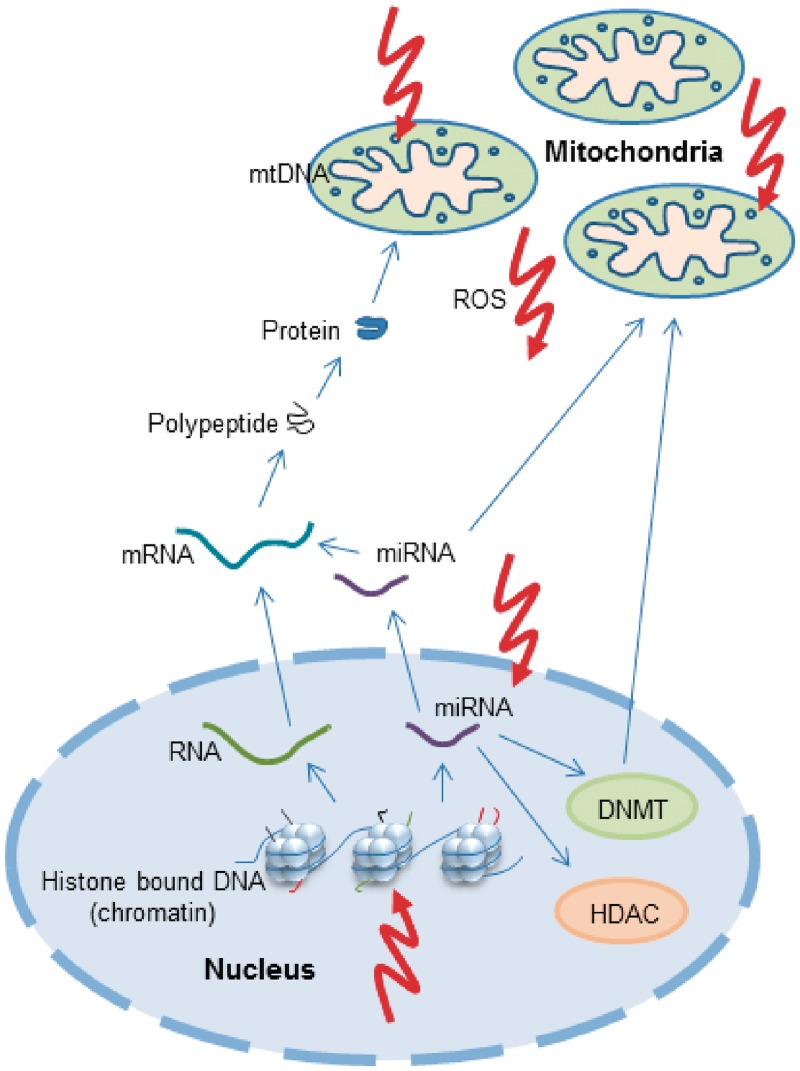
Over the last two decades, the mutation of mitochondrial DNA (mtDNA) has emerged as a major cause of inherited human disease. The disorders present clinically in at least 1 in 10,000 adults, but pathogenic mutations are found in approximately 1 in 200 of the background population. Mitochondrial DNA is maternally inherited and there can be marked phenotypic variability within the same family. Heteroplasmy is a significant factor and environmental toxins also appear to modulate the phenotype. Although genetic and biochemical studies have provided part of the explanation, a comprehensive understanding of the incomplete penetrance of these diseases is lacking--both at the population and family levels. Here, we review the potential role of epigenetic factors in the pathogenesis of mtDNA diseases and the contribution that epidemiological approaches can make to improve our understanding in this area. Despite being previously dismissed, there is an emerging evidence that mitochondria contain the machinery required to epigenetically modify mtDNA expression. In addition, the increased production of reactive oxygen species seen in several mtDNA diseases could lead to the epigenetic modification of the nuclear genome, including chromatin remodelling and alterations to DNA methylation and microRNA expression, thus contributing to the diverse pathophysiology observed in this group of diseases. These observations open the door to future studies investigating the role of mtDNA methylation in human disease.


Figures

Figure 1
Figure 1
A schematic representation of mitochondrial and nuclear genomes and their inter-relation with epigenetic factors. The nuclear genome is coiled around histone octamers to form nucleosomes. The tails of histone proteins are decorated with a variety of modifications that influence the regulation of gene expression. Permissive histone markings allow transcription from DNA to RNA, post-transcription processing to mRNA and translation to polypeptides and thus proteins. Nuclear-encoded mitochondrial proteins are then translocated into the mitochondrion. mtDNA also encodes genes essential for intra-mitochondrial protein synthesis, but this genome is not histone bound. In addition to mRNA, miRNA are also transcribed from nuclear DNA and can interfere with mRNA to induce degradation or suppress translation. miRNAs can influence mitochondrial metabolism and some miRNAs are known to directly activate the generation of ROS. Furthermore, miRNAs influence the expression of DNMT and HDAC enzymes. ROS produced by mitochondria or other endogenous sources can damage the mtDNA genome directly as well as influence epigenetic machinery at several levels, either through damage to miRNA or through the alteration of histone modifications. DNMTs translocate to the mitochondria and bind to mtDNA, although evidence that this is to effect epigenetic regulation remains elusive. HDAC, histone deacetylase; mRNA, messenger RNA; ROS, reactive oxygen species
Figure 2
Figure 2
The distribution of CpG sites in mtDNA. (a) The observed frequencies of the 16 dinucleotides divided by the expected frequencies for a random sequence based on the individual nucleotide frequencies. Values are plotted for the human mtDNA sequence plus 60 other mammalian species. (b) The density of the CpG sites in different sections of the human mtDNA sequence (rCRS), normalized by the average density of 0.026 per site. P-values are calculated by chi-square tests with Yates correction. OL and OH locations were taken from the mtDNA function locations list at MITOMAP (www.MitoMap.org) without alteration. OL was defined as 5721–5798 and OH was defined as 110–441
Figure 3
Figure 3
The location of CpG sites in the mitochondrial genome relative to mitochondrial haplogroup defining polymorphisms. The image shows the mitochondrial genome (centre), showing the position and relative size of each of the 13 major mitochondrial genes, the origins of heavy-strand and light-strand replication (OH and OL, respectively) as well as both the light-strand and heavy-strand promoter sites (PL and PH, respectively). Shown in the middle (in red) are the relative frequencies and positions of 3358 mtDNA variants (MAF = 0.01–49.6% of 2147 full European mtDNA sequences). The outer ring (in black) shows the relative position of each of the 435 predicted CpG sites
Figure 4
Figure 4
DNA methylation levels in nuclear-encoded mitochondrial genes in a control population. Twenty-four healthy female adult samples were analysed for genome-wide methylation using Illumina® HumanMethylation27 arrays. Probes were assigned to ‘non-mitochondrial’ or ‘mitochondrial’ groups as described in the main text. Histogram shows the DNA methylation distribution of CpG island probes assigned to non-mitochondrial or mitochondrial groups

References

    1. Wallace DC. Colloquium paper: bioenergetics, the origins of complexity, and the ascent of man. Proc Natl Acad Sci U S A. 2011;107(Suppl 2):8947–53. - PMC - PubMed
    1. Anderson S, Bankier AT, Barrell BG, et al. Sequence and organization of the human mitochondrial genome. Nature. 1981;290:457–65. - PubMed
    1. Smeitink J, van den Heuvel L, DiMauro S. The genetics and pathology of oxidative phosphorylation. Nat Rev Genet. 2001;2:342–52. - PubMed
    1. Zeviani M, Di Donato S. Mitochondrial disorders. Brain. 2004;127:2153–72. - PubMed
    1. Holt I, Harding AE, Morgan-Hughes JA. Deletion of muscle mitochondrial DNA in patients with mitochondrial myopathies. Nature. 1988;331:717–19. - PubMed

Publication types

Substances