

Disturbed cortico-subcortical interactions during motor task switching in traumatic brain injury
- PMID: 22287257
- PMCID: PMC6870055
- DOI: 10.1002/hbm.21508
Disturbed cortico-subcortical interactions during motor task switching in traumatic brain injury
Abstract
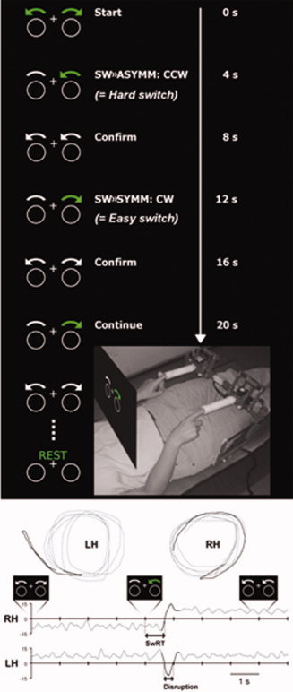
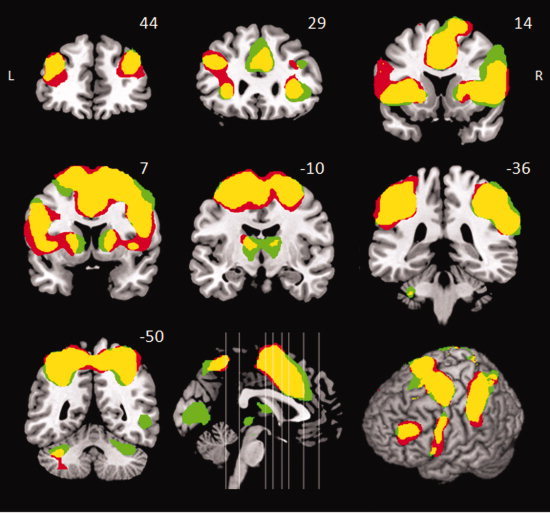
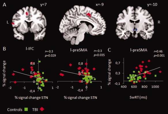
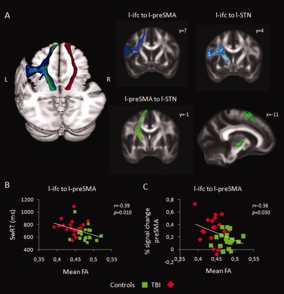
The ability to suppress and flexibly adapt motor behavior is a fundamental mechanism of cognitive control, which is impaired in traumatic brain injury (TBI). Here, we used a combination of functional magnetic resonance imaging and diffusion weighted imaging tractography to study changes in brain function and structure associated with motor switching performance in TBI. Twenty-three young adults with moderate-severe TBI and twenty-six healthy controls made spatially and temporally coupled bimanual circular movements. A visual cue signaled the right hand to switch or continue its circling direction. The time to initiate the switch (switch response time) was longer and more variable in the TBI group and TBI patients exhibited a higher incidence of complete contralateral (left hand) movement disruptions. Both groups activated the basal ganglia and a previously described network for task-set implementation, including the supplementary motor complex and bilateral inferior frontal cortex (IFC). Relative to controls, patients had significantly increased activation in the presupplementary motor area (preSMA) and left IFC, and showed underactivation of the subthalamic nucleus (STN) region. This altered functional engagement was related to the white matter microstructural properties of the tracts connecting preSMA, IFC, and STN. Both functional activity in preSMA, IFC, and STN, and the integrity of the connections between them were associated with behavioral performance across patients and controls. We suggest that damage to these key pathways within the motor switching network because of TBI, shifts the patients toward the lower end of the existing structure-function-behavior spectrum.
Copyright © 2012 Wiley Periodicals, Inc.
Figures


Similar articles
-
Reduced basal ganglia function when elderly switch between coordinated movement patterns.Cereb Cortex. 2010 Oct;20(10):2368-79. doi: 10.1093/cercor/bhp306. Epub 2010 Jan 15. Cereb Cortex. 2010. PMID: 20080932
-
Triangulating a cognitive control network using diffusion-weighted magnetic resonance imaging (MRI) and functional MRI.J Neurosci. 2007 Apr 4;27(14):3743-52. doi: 10.1523/JNEUROSCI.0519-07.2007. J Neurosci. 2007. PMID: 17409238 Free PMC article.
-
Task switching in traumatic brain injury relates to cortico-subcortical integrity.Hum Brain Mapp. 2014 May;35(5):2459-69. doi: 10.1002/hbm.22341. Epub 2013 Aug 2. Hum Brain Mapp. 2014. PMID: 23913872 Free PMC article.
-
Subcortical volume analysis in traumatic brain injury: the importance of the fronto-striato-thalamic circuit in task switching.Cortex. 2014 Feb;51:67-81. doi: 10.1016/j.cortex.2013.10.009. Epub 2013 Nov 4. Cortex. 2014. PMID: 24290948
-
Applications of Resting State Functional MR Imaging to Traumatic Brain Injury.Neuroimaging Clin N Am. 2017 Nov;27(4):685-696. doi: 10.1016/j.nic.2017.06.006. Epub 2017 Aug 18. Neuroimaging Clin N Am. 2017. PMID: 28985937 Free PMC article. Review.
Cited by
-
A Template and Probabilistic Atlas of the Human Sensorimotor Tracts using Diffusion MRI.Cereb Cortex. 2018 May 1;28(5):1685-1699. doi: 10.1093/cercor/bhx066. Cereb Cortex. 2018. PMID: 28334314 Free PMC article.
-
The subcortical cocktail problem; mixed signals from the subthalamic nucleus and substantia nigra.PLoS One. 2015 Mar 20;10(3):e0120572. doi: 10.1371/journal.pone.0120572. eCollection 2015. PLoS One. 2015. PMID: 25793883 Free PMC article.
-
Cortical Function in Acute Severe Traumatic Brain Injury and at Recovery: A Longitudinal fMRI Case Study.Brain Sci. 2020 Sep 3;10(9):604. doi: 10.3390/brainsci10090604. Brain Sci. 2020. PMID: 32899145 Free PMC article.
-
When the brain changes its mind: Oscillatory dynamics of conflict processing and response switching in a flanker task during alcohol challenge.PLoS One. 2018 Jan 12;13(1):e0191200. doi: 10.1371/journal.pone.0191200. eCollection 2018. PLoS One. 2018. PMID: 29329355 Free PMC article.
-
Cognitive and Motor Perseveration Are Associated in Older Adults.Front Aging Neurosci. 2021 Apr 27;13:610359. doi: 10.3389/fnagi.2021.610359. eCollection 2021. Front Aging Neurosci. 2021. PMID: 33986654 Free PMC article.
References
-
- Alexander MP, Stuss DT, Shallice T, Picton TW, Gillingham S ( 2005): Impaired concentration due to frontal lobe damage from two distinct lesion sites. Neurology 65: 572–579 - PubMed
-
- Amunts K, Schleicher A, Burgel U, Mohlberg H, Uylings HBM, Zilles K ( 1999): Broca's region revisited: Cytoarchitecture and intersubject variability. J Comp Neurol 412: 319–341 - PubMed
-
- Aravamuthan BR, Muthusamy KA, Stein JF, Aziz TZ, Johansen‐Berg H ( 2007): Topography of cortical and subcortical connections of the human pedunculopontine and subthalamic nuclei. Neuroimage 37: 694–705 - PubMed
-
- Aron AR ( 2007): The neural basis of inhibition in cognitive control. Neuroscientist 13: 214–228 - PubMed
Publication types
MeSH terms
LinkOut - more resources
Full Text Sources
