. 2013 Jun;34(6):1357-70.
doi: 10.1002/hbm.21515. Epub 2012 Jan 30.

Timing of posterior parahippocampal gyrus activity reveals multiple scene processing stages

Affiliations

Timing of posterior parahippocampal gyrus activity reveals multiple scene processing stages

Julien Bastin et al. Hum Brain Mapp. 2013 Jun.

Abstract

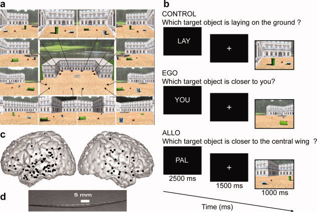
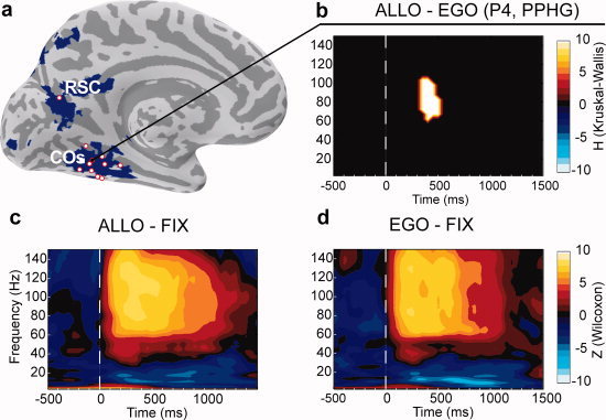
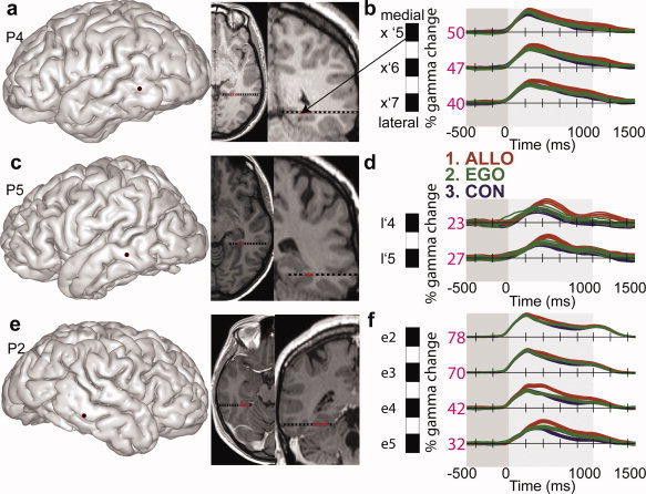
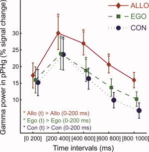
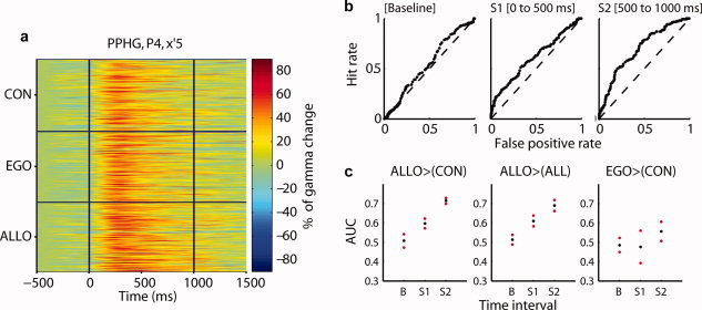
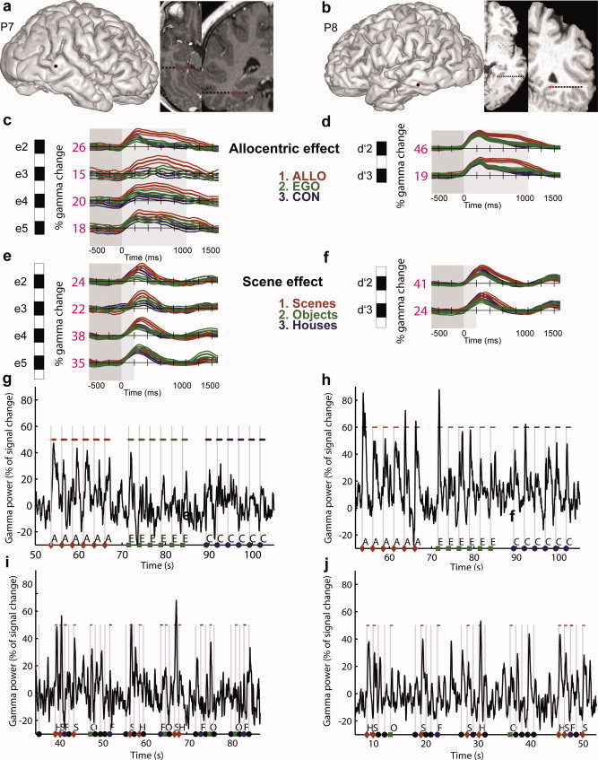
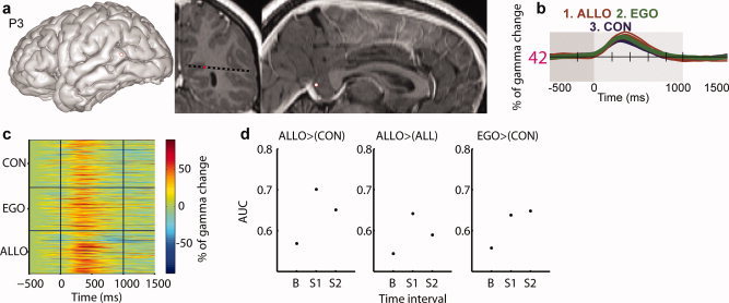
Posterior parahippocampal gyrus (PPHG) is strongly involved during scene recognition and spatial cognition. How PPHG electrophysiological activity could underlie these functions, and whether they share similar timing mechanisms is unknown. We addressed this question in two intracerebral experiments which revealed that PPHG neural activity dissociated an early stimulus-driven effect (>200 and <500 ms) and a late task-related effect (>600 and <800 ms). Strongest PPHG gamma band (50-150 Hz) activities were found early when subjects passively viewed scenes (scene selectivity effect) and lately when they had to estimate the position of an object relative to the environment (allocentric effect). Based on single trial analyses, we were able to predict when patients viewed scenes (compared to other visual categories) and when they performed allocentric judgments (compared to other spatial judgments). The anatomical location corresponding to the strongest effects was in the depth of the collateral sulcus. Our findings directly affect current theories of visual scene processing and spatial orientation by providing new timing constraints and by demonstrating the existence of separable information processing stages in the functionally defined parahippocampal place area.


Figures

Figure 1
Figure 1
Experimental design used to dissociate reference frame for spatial cognition (a and b) and entry points and structure of implanted intracerebral electrodes (c and d). (a) Viewpoints used to generate scenes stimuli. Objects positions within the scenes were independently manipulated relative to viewpoints. (b) Stimulus sequence. The main task was to judge which of the two target objects was closer to the patient's body/viewpoint (EGO) or which target object was closer to the central wing of the palace (ALLO). The control condition (CON) did not require position or distance estimation but required a shift of attention within the scene. The lower timeline shows the duration of each stimulus. (c) Electrodes entry point represented on a 3D reconstruction of the MNI brain. Each dot corresponds to a 1D array penetrating the brain orthogonally to the sagittal plane. (d) Multisites depth electrode. PAL, Palace (task instruction given before allocentric trials); YOU (task instruction given before egocentric trials); LAY, Laying (instruction given before control trials). [Color figure can be viewed in the online issue, which is available at wileyonlinelibrary.com.]
Figure 2
Figure 2
Typical pattern of oscillatory activity recorded from a PPHG site (patient P4, x′5, bd) and group PPHG sites anatomical location relative to brain areas specialized for allocentric coding as defined from a previous fMRI study (a). (a) Bold contrast Allo > Ego from a previous fMRI study (Committeri et al., 2004) is shown in blue on the inflated MNI brain. Electrode contacts from all patients located at the vicinity of the collateral sulcus or retrosplenial cortex are superimposed (red dots). (b) Typical allocentric statistical effect observed in an individual PPHG (Allo > Ego, Patient 4, KW test FDR corrected) was only observed in the gamma band (50–150 Hz). A positive H value (extracted from KW tests) corresponded to regions in the time‐frequency space where PPHG activity was stronger in the allocentric condition compared to the egocentric condition. (c and d) Time‐frequency representation of the electrophysiological responses recorded in the same PPHG site of Patient 4 (P4) in the Ego (c) and Allo (d) conditions. Warmer (yellow) colors correspond to a significant (Wilcoxon tests) power increase relative to baseline (−800 to 500 ms before stimulus), whereas cooler (blue) colors correspond to power significant power decreases. Wilcoxon tests were not corrected for multiple comparisons with FDR for illustrative purposes. [Color figure can be viewed in the online issue, which is available at wileyonlinelibrary.com.]
Figure 3
Figure 3
Reproducibility across patients of gamma‐band time courses as a function of spatial conditions in PPHG. (a, c, and e) Individual anatomical location of the electrode entry point on the patient's 3D brain is shown in addition to sagittal and coronal MRI slices (radiological convention) corresponding to the PPHG electrode contacts. (b, d, and f) Mean energy time courses (±95% confidence intervals) in the high gamma frequency band (50–150 Hz) is represented for all three conditions, expressed as a percentage of increase. The maximal value recorded for each electrode contact is also indicated (32). Lower case letters indicate different electrodes in each patient and the assigned numbers refer to the contact site within that electrode, with numbers increasing in the medial‐to‐lateral direction. Electrodes in the left hemisphere are indicated by (′) between the electrode letter and the contact site number (e.g., x′5–x′7 for patient P4), whereas right hemisphere electrodes are not (e.g., e2–e5 for patient P7). [Color figure can be viewed in the online issue, which is available at wileyonlinelibrary.com.]
Figure 4
Figure 4
Time course of the group averaged PPHG gamma activity (mean ± standard error) as a function of spatial conditions (n = 15 PPHG sites). Significance markers (*) reflect the results of the posthoc analysis performed to detail the time interval × spatial condition ANOVA interaction. For each spatial condition, the first time interval (0–200 ms) was compared to other time intervals to directly compare the duration of the gamma‐band increase in the allocentric, egocentric, and control conditions. The interaction corresponds to the fact that gamma activity was shorter in the control condition (< 400 ms), slightly longer in the egocentric condition (< 600 ms) and longest in the allocentric task (< 800 ms). [Color figure can be viewed in the online issue, which is available at wileyonlinelibrary.com.]
Figure 5
Figure 5
Selective single trial PPHG gamma amplitude increases in allocentric trials. (a) Example of single trial neural gamma‐band modulation expressed in percentage of change relative to baseline (P4, x′5). The electrode contact elicited a strong preference to allocentric processing. (b) Receiver operator characteristic (ROC) curves obtained by contrasting allocentric trials to all other trial types in three time intervals (same electrode as in a). (c) Average area under curves (AUC) extracted from ROC analyses in all PPHG sites (n = 15) and 95% confidence intervals. Allocentric trials could be readout when compared to control (left panel), to control and egocentric trials (middle panel), whereas egocentric trials were harder to discriminate although performance was above chance level (right panel). Baseline period, B = (−250 to 0 ms). Stimulus period S 1 = (0–500 ms); stimulus period S 2 = (500–1,000 ms poststimulus onset). [Color figure can be viewed in the online issue, which is available at wileyonlinelibrary.com.]
Figure 6
Figure 6
Gamma‐band responses in PPA dissociate two processing stages, across and within trials. (a and b) Anatomical location of PPA sites. (cf) Average (across trials) scene and allocentric effects in individual PPA sites (conventions are similar to Fig. 3). Scenes induced a stronger and longer gamma activity relative to other visual categories. This effect occurred and ended strikingly earlier than the allocentric effect (compare c–e and d–f panels). (gj) Raw single trials gamma‐band activity recorded in a typical PPA site (P7 e2 and P8, d′2) during the first (g and h) and second (i and j) experiment. The effect of allocentric coding (Allo > Ego) and scene encoding (Scene > Objects) can be directly seen within single trials gamma‐band activity. In Experiment 1, the highest peaks and longer gamma increases corresponded to the Allo judgments, whereas in Experiment 2, the highest peaks corresponded to scenes stimuli. A, allocentric; E, egocentric; C, control; S, scene; H, house; F, face; O, objects. [Color figure can be viewed in the online issue, which is available at wileyonlinelibrary.com.]
Figure 7
Figure 7
Retrosplenial cortex gamma‐band amplitude was highest in allocentric trials. (a) Anatomical location of RSC site (entry point and patient MRI slices). (b) Average gamma‐band time course as a function of experimental conditions. (c) Single trial gamma‐band amplitude (in percentage of signal change). (d) RSC readout index values (ROC area under curve, AUC). Figure conventions are similar to Figures 3 and 5. [Color figure can be viewed in the online issue, which is available at wileyonlinelibrary.com.]

Similar articles

Cited by

References

    1. Aguirre GK, D'Esposito M ( 1999): Topographical disorientation: A synthesis and taxonomy. Brain 122( Pt 9): 1613–1628. - PubMed
    1. Aguirre GK, Zarahn E, D'Esposito M ( 1998): An area within human ventral cortex sensitive to “building” stimuli: Evidence and implications. Neuron 21: 373–383. - PubMed
    1. Bar M ( 2004): Visual objects in context. Nat Rev Neurosci 5: 617–629. - PubMed
    1. Boccara CN, Sargolini F, Thoresen VH, Solstad T, Witter MP, Moser EI, Moser MB ( 2010): Grid cells in pre‐ and parasubiculum. Nat Neurosci 13: 987–994. - PubMed
    1. Bohbot VD, Kalina M, Stepankova K, Spackova N, Petrides M, Nadel L ( 1998): Spatial memory deficits in patients with lesions to the right hippocampus and to the right parahippocampal cortex. Neuropsychologia 36: 1217–1238. - PubMed

Publication types