. 2012 Dec;34(12):1734-46.
doi: 10.1002/hed.22004. Epub 2012 Jan 27.

Induction of MAGE-A3 and HPV-16 immunity by Trojan vaccines in patients with head and neck carcinoma

Affiliations

Induction of MAGE-A3 and HPV-16 immunity by Trojan vaccines in patients with head and neck carcinoma

Caroline J Voskens et al. Head Neck. 2012 Dec.

Abstract

Background: We performed a pilot study using Trojan vaccines in patients with advanced squamous cell carcinoma of the head and neck (SCCHN). These vaccines are composed of HLA-I and HLA-II restricted melanoma antigen E (MAGE)-A3 or human papillomavirus (HPV)-16 derived peptides, joined by furin-cleavable linkers, and linked to a "penetrin" peptide sequence derived from HIV-TAT. Thirty-one patients with SCCHN were screened for the trial and 5 were enrolled.

Methods: Enrolled patients were treated with 300 μg of Trojan peptide supplemented with Montanide and granulocyte-macrophage colony-stimulating factor (GM-CSF) at 4-week intervals for up to 4 injections.

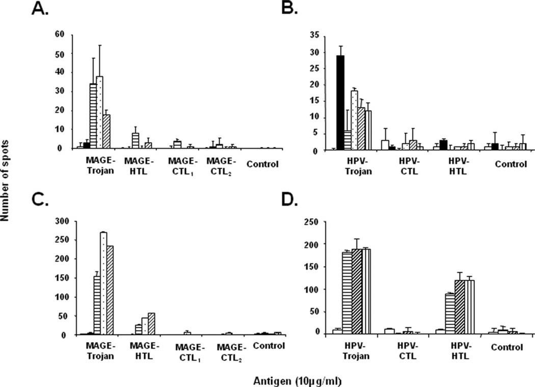
Results: Following vaccination, peripheral blood mononuclear cells (PBMCs) from 4 of 5 patients recognized both the full Trojan constructs and constituent HLA-II peptides, whereas responses to HLA-I restricted peptides were less pronounced.

Conclusion: This treatment regimen seems to have acceptable toxicity and elicits measurable systemic immune responses against HLA-II restricted epitopes in a subset of patients with advanced SCCHN.


Conflict of interest statement

Conflict of interest: Dr. Strome is a major stockholder in Gliknik, a biotechnology company. Gliknik has licensed the rights to this vaccine technology from the University of Maryland. Dr. Strome receives royalties from the licensure of this technology. Dr. Celis receives royalties from the Mayo Clinic College of Medicine as an inventor of the Trojan peptide vaccines.

Figures

FIGURE 1
FIGURE 1
Immunization schedule. All enrolled patients were immunized subcutaneously in the inguinal region at 4-week intervals for up to 4 injections. After the final immunization, patients returned for follow-up visits every 3 months.
FIGURE 2
FIGURE 2
The T2-weighted brain MR images of patient 1 and Trojan vaccine-specific immunity. Left image (A) obtained 8 weeks before vaccination (January 30, 2006), middle image (B) obtained during neurological event (April 21, 2006), and right image (C) obtained after resolution of neurological symptoms (July 14, 2006). Arrows indicate the site of the cerebral metastasis. Peripheral blood mononuclear cells (PBMCs) were restimulated once with melanoma antigen E (MAGE)-Trojan vaccine and cultured in the presence of interleukin (IL)-7 (5 ng/ mL) and IL-15 (5 ng/mL). After 7 days, cells were evaluated for MAGE Trojan-specific HLA-I and HLA-II peptide (D) specificity by interferon-γ (IFN-γ) Elispot assay. Number of spots per 100,000 PBMCs is shown.
FIGURE 3
FIGURE 3
Trojan peptide-based vaccines induce Trojan-specific precursors in vivo. Peripheral blood mononuclear cells (PBMCs) collected before immunization and at indicated time points after immunization were evaluated for Trojan specificity by interferon-γ (IFN-γ) Elispot assay. Direct ex vivo measurement of T-cell reactivity against melanoma antigen E (MAGE)-Trojan (A) human papillomavirus (HPV)-Trojan (B) and individual Trojan-specific HLA-I and HLA-II peptides (A and B). Number of spots per 200,000 PBMCs is shown. Error bars represent SD from triplicate wells. Control values are shown without subtraction from experimental values. PBMCs were restimulated once with MAGE-Trojan or HPV-Trojan vaccine and cultured in the presence of interleukin (IL)-7 (5 ng/mL) and IL-15 (5 ng/mL). After 7 days, cells were evaluated for MAGE-Trojan (C), HPV-Trojan (D), and individual Trojan-specific HLA-I and HLA-II peptide (C and D) specificity by IFN-γ Elispot assay. Number of spots per 100,000 PBMC is shown. Error bars represent SD from triplicate wells. * p < .05.
FIGURE 4
FIGURE 4
Vaccination with melanoma antigen E (MAGE)-A3 and human papillomavirus (HPV)-16 specific Trojan vaccines elicits immunoglobulin G (IgG) antibody responses in patients with squamous cell carcinoma of the head and neck (SCCHN). Trojan-specific IgG was quantified in plasma obtained before immunization and at various time-points after immunization as indicated. MAGE-A3 Trojan (A and B) and HPV Trojan (C and D) specific IgG was quantified by enzyme-linked immunosorbent assay (ELISA). Data represent mean optical density (OD) readings from triplicate wells derived from 3 different plasma dilutions.
FIGURE 5
FIGURE 5
Trojan-specific precursor frequency at the injection site exceeds precursor frequency in the peripheral circulation. Frozen section slides were generated from a granuloma derived from the site of immunization and stained by immunohistochemistry with a variety of leukocyte-specific markers. The granuloma was biopsied 19 days after the second vaccination. Positron emission tomography (PET) scan revealing high levels of inflammation at the injection sites as indicated by arrows (A); high numbers of antigen-presenting cells (CD68+) and CD3+ T cells infiltrated the site of immunization (B); part of the granuloma was enzymatically digested into a single cell suspension and evaluated for Trojan vaccine-specificity (C). As measured by flow cytometry, the majority of CD3+ T cells in the granuloma were found to be CD4+ cells. Next, cells were directly ex vivo evaluated for Trojan-specificity by interferon-γ (IFN-γ) Elispot. High numbers of Trojan-specific cells were found at the site of injection (D). In contrast, at the same time interval, no Trojan-specific responses were detected in cells peripheral blood mononuclear cells (PBMCs) derived from the peripheral circulation. Number of spots per 50,000 cells (granuloma) and 100,000 PBMCs is shown. Error bars represent SD from triplicate wells. * p < .05. [Color figure can be viewed in the online issue, which is available at wileyonlinelibrary.com.]
FIGURE 6
FIGURE 6
The CD8+ and CD4+ T cells infiltrate the tumor microenvironment during immunization and a subset appears to be human papillomavirus (HPV)-specific. Photomicrographs of immunohistochemical staining of tumor tissue harvested before and after immunization with HPV-Trojan vaccine. High numbers of CD8+ and CD4+ cells were found in the tumor microenvironment after immunization (A). Representative sections from CD4 and CD8 staining of the neck metastasis slides before and 3 months after the last vaccination are shown. Positive controls were positive for all experiments (inset CD4). Part of the neck metastasis, collected at 3 months after immunization, was enzymatically digested into a single cell suspension tumor infiltrating lymphocytes (TILs). Peripheral blood mononuclear cells (PBMCs) collected before and PBMCs and TILs collected after 3 months of immunization were directly ex vivo stained with HPV-16 specific HLA-A*0201 tetramer and analyzed by flow cytometry for Trojan HLA-I specificity (B). In comparison to PBMCs collected at the same time-interval, TILs derived from the tumor microenvironment after 3 months of immunization were found to be HPV-16 specific, 0.4% and 8.1%, respectively. [Color figure can be viewed in the online issue, which is available at wileyonlinelibrary.com.]

References

    1. Ahmed SM, Cohen EE. Treatment of squamous cell carcinoma of the head and neck in the metastatic and refractory settings: advances in chemotherapy and the emergence of small molecule epidermal growth factor receptor kinase inhibitors. Curr Cancer Drug Targets. 2007;7:666–673. - PubMed
    1. Voskens CJ, Strome SE, Sewell DA. Synthetic peptide-based cancer vaccines: lessons learned and hurdles to overcome. Curr Mol Med. 2009;9:683–693. - PubMed
    1. Rosenberg SA, Yang JC, Restifo NP. Cancer immunotherapy: moving beyond current vaccines. Nat Med. 2004;10:909–915. - PMC - PubMed
    1. Zwaveling S, Ferreira Mota SC, Nouta J, et al. Established human papillomavirus type 16-expressing tumors are effectively eradicated following vaccination with long peptides. J Immunol. 2002;169:350–358. - PubMed
    1. Hoffmann TK, Bier H, Whiteside TL. Targeting the immune system: novel therapeutic approaches in squamous cell carcinoma of the head and neck. Cancer Immunol Immunother. 2004;53:1055–1067. - PMC - PubMed

Publication types

MeSH terms