. 2012 Feb;40(4):1868-78.
doi: 10.1093/nar/gks022. Epub 2012 Jan 27.

A human XRCC4-XLF complex bridges DNA

Affiliations

A human XRCC4-XLF complex bridges DNA

Sara N Andres et al. Nucleic Acids Res. 2012 Feb.

Abstract

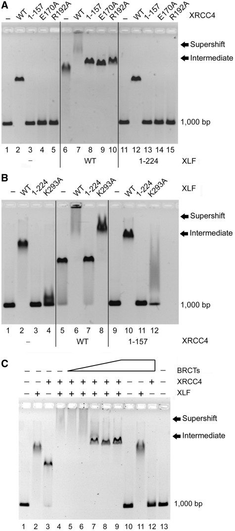
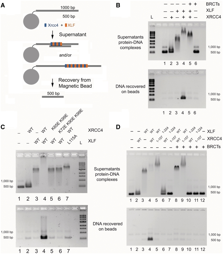
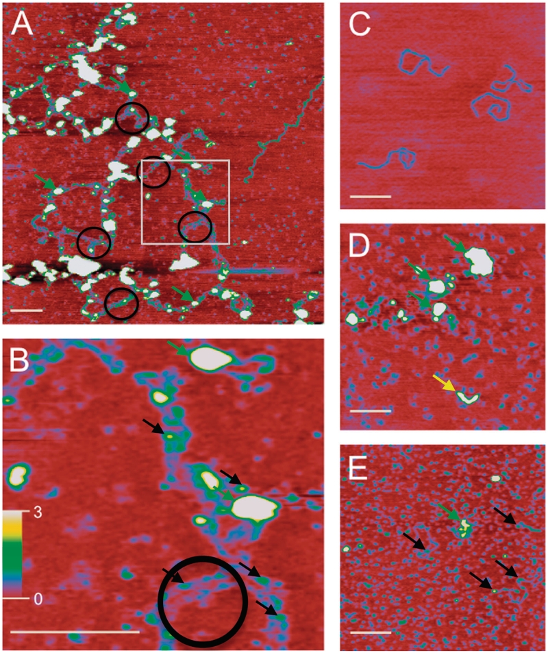
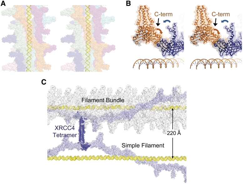
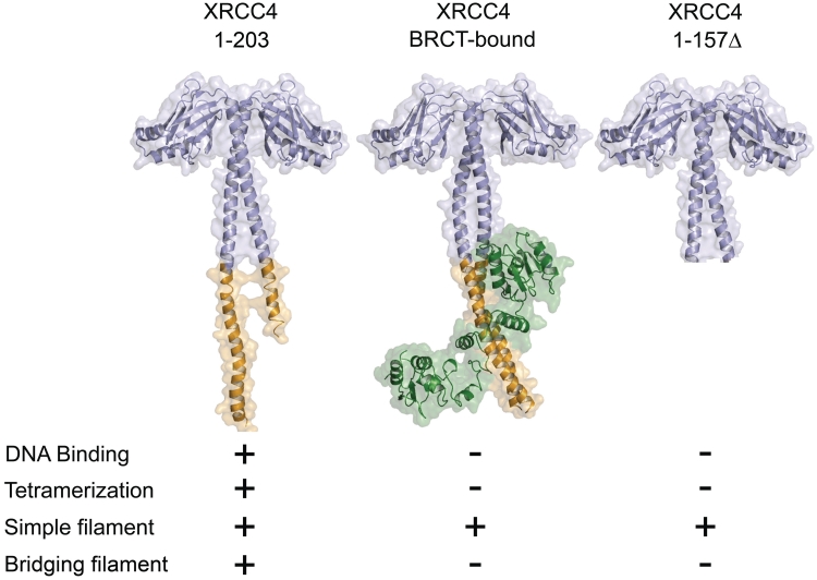
DNA double-strand breaks pose a significant threat to cell survival and must be repaired. In higher eukaryotes, such damage is repaired efficiently by non-homologous end joining (NHEJ). Within this pathway, XRCC4 and XLF fulfill key roles required for end joining. Using DNA-binding and -bridging assays, combined with direct visualization, we present evidence for how XRCC4-XLF complexes robustly bridge DNA molecules. This unanticipated, DNA Ligase IV-independent bridging activity by XRCC4-XLF suggests an early role for this complex during end joining, in addition to its more well-established later functions. Mutational analysis of the XRCC4-XLF C-terminal tail regions further identifies specialized functions in complex formation and interaction with DNA and DNA Ligase IV. Based on these data and the crystal structure of an extended protein filament of XRCC4-XLF at 3.94 Å, a model for XRCC4-XLF complex function in NHEJ is presented.


Figures

Figure 1.
Figure 1.
XRCC4–XLF DNA binding. (A) XRCC4 wild-type (WT) and mutants (8 µM) were incubated with 100 ng of DNA with or without 2 µM XLF (WT and mutants), analyzed by EMSA. (B) WT XLF and its mutants (2 µM) were incubated with 100 ng of DNA with or without 8 µM XRCC4 (WT and mutants). (C) Effect of DNA Ligase IV tandem BRCT domains on XRCC4–XLF–DNA complex formation. XRCC4 (8 µM) and XLF (2 µM) were incubated with 100 ng DNA fragments in the presence of increasing amounts of DNA Ligase IV tandem BRCT domains (BRCTs, 0, 0.5, 1, 2, 4–8 µM).
Figure 2.
Figure 2.
Bridging of DNA molecules by XRCC4–XLF. (A) Schematic of DNA-bridging assay. Proteins were incubated with magnetic beads linked to 1000-bp DNA and free 500-bp DNA. Beads were separated from supernatant and analyzed separately for presence of the 500-bp DNA. (B) an amount of 200 ng each of 1000- and 500-bp DNA fragments were incubated with XRCC4 (2 µM), XLF (2 µM) or DNA Ligase IV tandem BRCT domains (BRCTs, 2 µM). Top panel shows the analysis of the protein–DNA complexes in the supernatants. Bottom panel shows the recovery of DNA species on the beads. L = 1 kb DNA ladder (NEB). (C) Bridging assays performed as in (B) with mutants preventing XRCC4–XLF filament formation. (D) Bridging assays performed as in (B). XRCC41–157 and XLF1–224 are truncated proteins lacking C-terminal tails and DNA-binding activity.
Figure 3.
Figure 3.
SFM analysis of protein–DNA networks. (A) Large protein–DNA networks observed in XRCC4–XLF DNA-binding reactions. Nucleoprotein complexes appear as higher wider objects (∼30–60 Å high and 400–800 Å wide, green arrows). Such complexes were typical and observed on all 16 of the 2 × 2 micron images collected for this sample. Protein-induced parallel bridging of DNA molecules is evident (black circles) and occurs in many places along the long complex. Extended DNA networks indicate DNA molecules are positioned end-to-end. White box indicates area enlarged in (B). (B) Enlargement of (A) highlighting parallel DNA molecules. Spacing between parallel DNA molecules is ∼200 Å. Smaller nucleoprotein complexes, measuring 4–7 Å high and 110–180 Å wide, are also evident (Figure 3B, black arrows). (C) The 1.8-kb linear DNA, contour length 6000 Å, width 180 Å and height 2 Å. (D) Image of XRCC4–XLF complexes in absence of DNA. Small protein complexes, likely dimers, tetramers and small multimers, appear as blue uniform objects distributed over the surface. Larger protein complexes (30–60 Å high and 400–800 Å wide) and elongated forms were also present and are indicated by green and yellow arrows, respectively. Elongated structures (yellow arrow) measuring ∼1300 Å long, 20–25 Å high and 300 Å wide, were observed with a frequency of approximately one for every 2 × 2 micron image inspected. (E) Addition of DNA Ligase IV BRCT domains disrupts nucleoprotein networks. Smaller protein complexes similar to (D) are uniformly distributed and appear as small blue objects. DNA molecules associated with protein complexes are also observed (25–60 Å high and 400–800 Å wide, green arrow) with a frequency of approximately one for every 2 × 2 micron image inspected. Smaller nucleoprotein complexes (4–7 Å high and 110–180 Å wide, black arrows) were observed at higher frequency. (A) is 2 × 2 microns. (B) is 500 × 500 nm. (C–E) are 1 × 1 microns. In all images the white bar is 2000 Å long and height is indicated by color (0–3 nm red to yellow/white, scale bar in panel B). Note that biomolecule dimensions are distorted in SFM images. X–Y dimensions increase due to tip convolution and Z (height) decreases relative to tip surface and tip molecule interactions. For instance DNA, that is 20 Å wide and 20 Å high based on crystal structure of B-form, typically measures 200 Å wide and 2–5 Å high in our SFM images. Relative size and separation between objects can be used very accurately.
Figure 4.
Figure 4.
Crystal structure of the XRCC41–157–XLF1–224 complex. (A) Interaction of XLF1–224 homodimer (orange) with an adjacent XRCC41–157 homodimer (blue) as seen in the crystal structure. Black arrow illustrates ∼30° offset between homodimers. (B) XRCC41–157–XLF1–224 filament. XRCC41–157 homodimers are numbered. Colored arrows indicate directionality of XRCC41–157 (blue) and XLF1–224 (orange) C-terminal tails. Filament diameter is indicated in Å. (C) XRCC41–157–XLF1–224 filament, rotated 90° clockwise from (B). Length of one filament revolution is indicated in Å. (D) XRCC41–157–XLF1–224 head-to-head interface. (E) Key amino acids directly involved in XRCC41–157–XLF1–224 head-to-head interaction.
Figure 5.
Figure 5.
Model of XRCC4–XLF filaments bound to DNA. (A) Multiple adjacent filaments bound to DNA (yellow). Each color is a separate filament. DNA runs through the pore of the XRCC4–XLF filament. (B) Tails of an XRCC41–157 homodimer (blue) point toward the N-terminus of XLF1–224 (orange), in an adjacent filament. (C) XRCC4 dimers associate into a tetramer through C-terminal tails (PDB 1FU1). DNA-bridging models illustrating single and complex filaments. Two DNA molecules coated in a simple or multi-filament bundle are bridged through XRCC4 C-terminal tails.
Figure 6.
Figure 6.
Summary of the structural states of XRCC4. Structural states of XRCC4 are indicated with their associated function (PDB 1FU1 and PDB 3II6; 21,23,37,38).

Comment on

References

    1. Kuzminov A. Single-strand interruptions in replicating chromosomes cause double-strand breaks. Proc. Natl Acad. Sci. USA. 2001;98:8241–8246. - PMC - PubMed
    1. Roth DB, Nakagima PB, Menetski JP, Bosma MJ, Gellert M. V(D)J recombination in mouse thymocytes: double-strand breaks near T cell receptor delta rearrangement signals. Cell. 1992;69:41–53. - PubMed
    1. Gottlieb TM, Jackson SP. The DNA-dependent protein kinase: requirement for DNA ends and association with Ku antigen. Cell. 1993;72:131–142. - PubMed
    1. Ding Q, Reddy YV, Wang W, Woods T, Douglas P, Ramsden DA, Lees-Miller SP, Meek K. Autophosphorylation of the catalytic subunit of the DNA-dependent protein kinase is required for efficient end processing during DNA double-strand break repair. Mol. Cell Biol. 2003;23:5836–5848. - PMC - PubMed
    1. McElhinny SA, Havener JM, Garcia-Diaz M, Juárez R, Bebenek K, Kee BL, Blanco L, Kunkel TA, Ramsden DA. A gradient of template dependence defines distinct biological roles for family × polymerases in nonhomologous end joining. Mol. Cell. 2005;19:357–366. - PubMed

Publication types