. 2013 Jan;62(1):83-93.
doi: 10.1136/gutjnl-2011-301601. Epub 2012 Jan 27.

A basal gradient of Wnt and stem-cell number influences regional tumour distribution in human and mouse intestinal tracts

Affiliations
Free PMC article

A basal gradient of Wnt and stem-cell number influences regional tumour distribution in human and mouse intestinal tracts

Simon J Leedham et al. Gut. 2013 Jan.
Free PMC article

Abstract

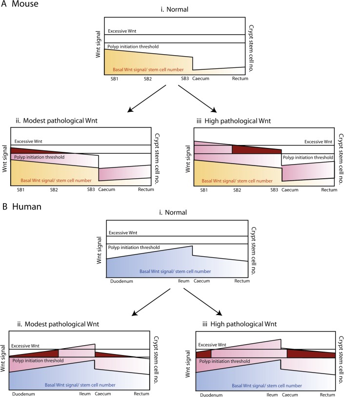
Objective: Wnt signalling is critical for normal intestinal development and homeostasis. Wnt dysregulation occurs in almost all human and murine intestinal tumours and an optimal but not excessive level of Wnt activation is considered favourable for tumourigenesis. The authors assessed effects of pan-intestinal Wnt activation on tissue homeostasis, taking into account underlying physiological Wnt activity and stem-cell number in each region of the bowel.

Design: The authors generated mice that expressed temporally controlled, stabilised β-catenin along the crypt-villus axis throughout the intestines. Physiological Wnt target gene activity was assessed in different regions of normal mouse and human tissue. Human intestinal tumour mutation spectra were analysed.

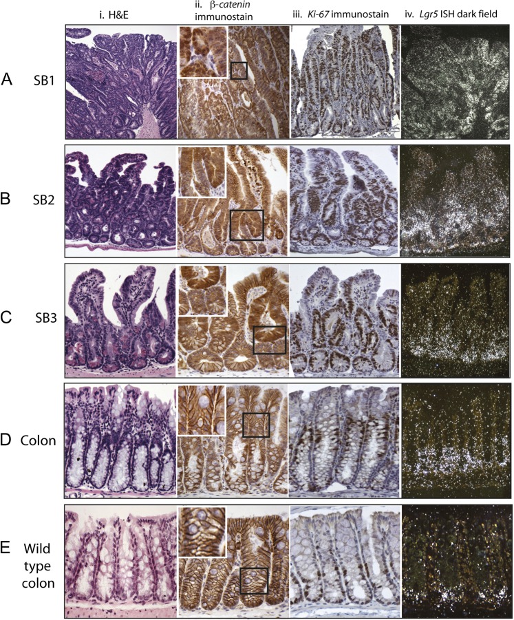
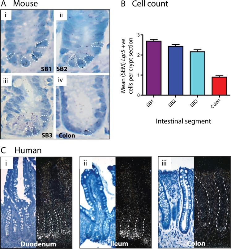
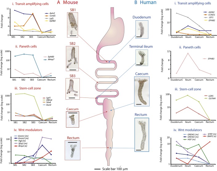
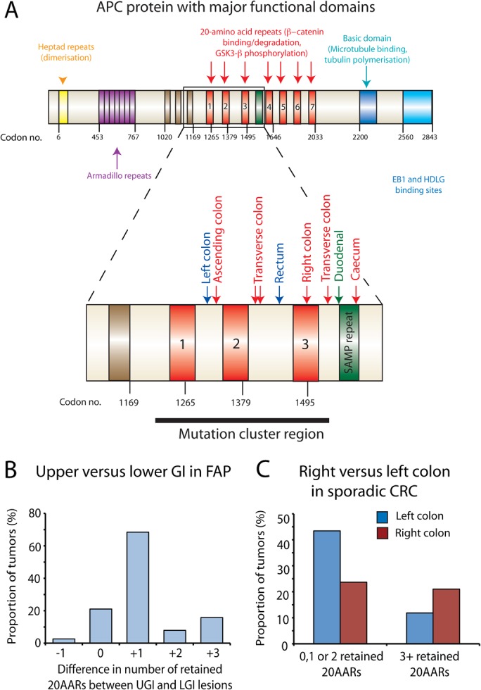
Results: In the mouse, β-catenin stabilisation resulted in a graduated neoplastic response, ranging from dysplastic transformation of the entire epithelium in the proximal small bowel to slightly enlarged crypts of non-dysplastic morphology in the colorectum. In contrast, stem and proliferating cell numbers were increased in all intestinal regions. In the normal mouse and human intestines, stem-cell and Wnt gradients were non-identical, but higher in the small bowel than large bowel in both species. There was also variation in the expression of some Wnt modulators. Human tumour analysis confirmed that different APC mutation spectra are selected in different regions of the bowel.

Conclusions: There are variable gradients in stem-cell number, physiological Wnt activity and response to pathologically increased Wnt signalling along the crypt-villus axis and throughout the length of the intestinal tract. The authors propose that this variation influences regional mutation spectra, tumour susceptibility and lesion distribution in mice and humans.


Conflict of interest statement

Competing interests: None.

Figures

Figure 1
Figure 1
Stabilised β-catenin mouse dysplastic change in the small intestine parallels expansion of proliferating cells with abnormal β-catenin expression. In all intestinal regions there was expansion of the mid-crypt zone. In the proximal small bowel (SB1) this was so pronounced that it destroyed the crypt-villus architecture (Ai). The expanded mid-crypt cell population was dysplastic, actively proliferating (Ki-67+ immunostaining) (Aiii) and had abnormal expression of β-catenin (heavy nuclear or cytoplasmic) (Aii). Distal small bowel (SB3) to mid-small bowel (SB2) there was some preservation of normal cell morphology at the crypt bases and villi tips with Ki-67 negative cells (B, Ciii) and membranous β-catenin stain (B, Cii). The entire colon was morphologically non-dysplastic with membranous β-catenin staining (Dii); however, there was an expansion and upward shift of the Ki-67+ cells in comparison with wild-type controls (Diii). Expansion of the stem-cell zone using S35-UTP labelled in situ hybridisation for Lgr5 (A–Eiv). There was expansion in the number of Lgr5 expressing cells in all intestinal regions. In the Ctnnb1Δex3 mouse colon the Lgr5+ cells were restricted to crypt bases but filled the bottom third of the phenotypically non-dysplastic crypt (Div), a considerable expansion of the stem-cell compartment in comparison with wild-type mouse colon (Eiv).
Figure 2
Figure 2
Stem-cell numbers vary through the intestinal tract. (A) Mouse in situ hybridisation (ISH). H3-UTP labelled ISH for Lgr5 on well-orientated crypts from a wild-type mouse: (i) proximal small bowel (SB1); (ii) mid-small bowel (SB2); (iii) distal small bowel (SB3); and (iv) colon. Individual cells containing ≥4 silver granules were counted and are highlighted in each picture. (B) Mouse Lgr5+ cell counts. Mean (SEM) Lgr5+ cell counts are shown. Lgr5+ cells are maximal in the SB1 with a slightly reducing number through to the ileum (SB3). A profound drop in Lgr5+ cells is seen in the colon (p<0.001, analysis of variance). Cell numbers were counted in 100 crypts from each region from three different wild-type mice. (C) Human ISH. S35-UTP labelled ISH for LGR5 on human endoscopic biopsy specimens from: (i) duodenum (ii) ileum and (iii) colon of the same patient. LGR5 expression was restricted to the crypt bases and was maximal in the ileum, with only single LGR5+ cells seen in colonic crypts. ISH was carried out on biopsy sections from 7 different patients undergoing normal upper and lower endoscopies.
Figure 3
Figure 3
Individual crypt Wnt target gene expression varies longitudinally throughout the normal mouse and human intestinal tract. Thumbnail pictures show whole mount intestinal crypts and villi from the different regions of normal mouse and human intestine after EDTA extraction. Mouse villi are pictured purely to provide scale; in fact, only individual crypts were aspirated and used in analysis. Mesenchymal tissue was isolated after complete epithelial denudement. Scale bar 100 μm. Five crypts were dissected from each intestinal region, analysed individually and used to calculate a mean ΔCt, which was then averaged across three wild-type mice (online supplementary table 2A) and four human patients (online supplementary table 2B). Mean ΔΔCt values were used to calculate expression fold changes compared with the region of minimal expression. (A) Mouse gene expression category 1 Wnt target genes (transit amplifying cells) showed quite variable expression along the intestine but were maximally expressed in the proximal small bowel (SB1) (with the exception of Cyclin D1). Category 2 genes (Paneth cells) were expressed in the small intestine as expected from this cell distribution (p<0.001, analysis of variance (ANOVA)). Category 3 genes (stem-cell zone) showed very similar expression profiles with maximal expression in the small intestine declining markedly in the colon with minimal expression seen in the caecum (p<0.001, ANOVA). Sox4 expression closely mirrored that of Ascl2 and a striped line indicates the regions of identical expression. Conversely, epithelial Bmp2 expression was minimal in the duodenum climbing steadily to a peak in the mouse rectum (p<0.001, ANOVA), and there was also significant variability in the expression of some of the mesenchymal Wnt modulators (Grem2 and Hgf, p<0.05, ANOVA). The expression of the mesenchymal Wnt antagonist, Sfrp2, varied considerably with colonic expression more than 130-fold greater than that in the small intestine (p=0.035, ANOVA). (B) Human gene expression. All human Wnt target genes were maximally expressed in the ileum and minimally expressed in the rectum with the exception of the category 1 genes LEF1 and CYCLIN D1 (CCND1), which were maximally expressed more proximally. Category 3 (stem-cell) gene expression peaked in the ileum correlating with the human in situ findings (p<0.001, ANOVA). Caecal stem-cell marker expression is significantly greater than that seen in the rectum. The inverse correlation was seen for epithelial BMP2 expression (p<0.001, ANOVA). Expression of the mesenchymal Wnt modulators was generally higher in the human small intestine which contrasted sharply with the murine Gremlin 1, 2 and Sfrp2 expression gradients. Only the expression gradient for HGF reached statistical significance in the humans (p=0.05, ANOVA) as the result of a 10-fold difference in expression from the caecum to the rectum. SB1, proximal small bowel; SB2, mid-small bowel; SB3, distal small bowel.
Figure 4
Figure 4
The adenomatous polyposis coli (APC) mutation spectrum varies with intestinal region in familial adenomatous polyposis (FAP) and sporadic gastrointestinal tumours. (A) Mutation spectra of upper and lower gastrointestinal (GI) lesions taken from the same FAP patient. The major functional domains of the APC protein are shown with an enlargement of the mutation cluster region. The number of retained 20-amino acid β-catenin binding repeats (20AARs) was calculated in upper GI (UGI) and lower GI (LGI) lesions taken from the same individual FAP patient(s) after determination of the mutation spectra of each lesion. The mutations for one individual patient (patient 1, online supplementary table 3) are mapped out (UGI lesion, green; right colon, red; left colon, blue) (B). UGI lesions in FAP retain significantly more 20AARs than LGI lesions taken from the same patient. The number of retained repeats from LGI lesions was subtracted from the number of repeats retained in the same patients' UGI lesions and the difference plotted. Significantly more repeats were retained in UGI lesions resulting in a more modest Wnt perturbation in these lesions (p<0.001, binomial test). (C) Right sided sporadic colorectal cancers and cell lines retain significantly more 20AARs than left sided lesions. Leisons in our sporadic data set of 76 tumours (online supplementary table 4A and B) were categorised by their distribution and the cumulative number of retained 20AARs after identification of first and second hits at APC in all lesions. Tumours retaining a cumulative total of three or more 20AARs were more likely to arise proximally whereas on the left side of the colon, mutations that retain fewer 20AARs were optimally selected to produce a greater Wnt perturbation in the resultant polyp (p=0.03, Fishers Exact test).
Figure 5
Figure 5
‘Just-right’ theory and genotype–phenotype correlations. (Ai and Bi) Basal Wnt signalling and stem-cell number in the mouse and humans. Mouse and human basal Wnt target expression profile and stem-cell number vary down the length of intestinal tract (figure 3). The two species have quite distinct profiles which in combination with ‘just-right’ mutation spectra may contribute to intestinal lesion distribution. (Aii and Aiii) The consequences of pathological Wnt signalling in the mouse. Submaximal pathological Wnt perturbation (seen in Ctnnb1Δex3 and Apc1322T mice, as evidenced by low levels of nuclear β-catenin expression14) breaches the dysplasia threshold in the proximal small intestine where basal Wnt expression and stem-cell numbers are the highest, resulting in a heavy proximal small bowel (SB1) and mid-small bowel (SB2) lesion burden. In the mouse colon, the Wnt perturbation is insufficient to cause dysplasia in the colon despite causing an increase in the stem-cell number. Maximal Wnt perturbation as seen in the ApcMin(R850X) is excessive and thus suboptimal proximally; hence, a predominantly distal small bowel (SB3) polyp distribution is characteristic. Maximal lesion distribution shown with dark red shading. (Bii and Biii) The consequences of pathological Wnt signalling in the humans. Modest Wnt perturbations such as that resulting from mutations that retain several 20-amino acid β-catenin binding repeats (20AARs) favour lesion development in the upper gastrointestinal (GI) and right colonic regions, explaining the more severe upper GI phenotype seen in patients with germline mutations after codon 1400. Conversely, early mutation cluster region mutations retain fewer 20AARs resulting in a greater degree of Wnt perturbation. Excessive Wnt is suboptimal for upper GI lesions but favours more distal lesions resulting in a severe colonic phenotype with loss of heterozygosity optimally selected in the rectum of patients with germline codon 1309 mutations. Maximal lesion distribution is shown with dark red shading.

References

    1. Scoville D, Sato T, He X, et al. Current view: intestinal stem cells and signaling. Gastroenterology 2008;134:849–64 - PubMed
    1. Kuhnert F, Davis CR, Wang HT, et al. Essential requirement for Wnt signaling in proliferation of adult small intestine and colon revealed by adenoviral expression of Dickkopf-1. Proc Natl Acad Sci U S A 2004;101:266–71 - PMC - PubMed
    1. Radtke F, Clevers H, Riccio O. From gut homeostasis to cancer. Curr Mol Med 2006;6:275–89 - PubMed
    1. Sieber OM, Tomlinson IP, Lamlum H. The adenomatous polyposis coli (APC) tumour suppressor—genetics, function and disease. Mol Med Today 2000;6:462–9 - PubMed
    1. Powell SM, Zilz N, Beazer-Barclay Y, et al. APC mutations occur early during colorectal tumorigenesis. Nature 1992;359:235–7 - PubMed

Publication types

MeSH terms