. 2012 Apr 15;302(8):L775-84.
doi: 10.1152/ajplung.00196.2011. Epub 2012 Jan 27.

Vasculoprotective effects of heme oxygenase-1 in a murine model of hyperoxia-induced bronchopulmonary dysplasia

Affiliations

Vasculoprotective effects of heme oxygenase-1 in a murine model of hyperoxia-induced bronchopulmonary dysplasia

Angeles Fernandez-Gonzalez et al. Am J Physiol Lung Cell Mol Physiol. .

Abstract

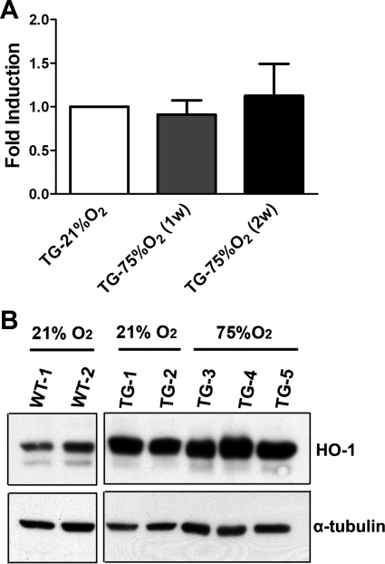
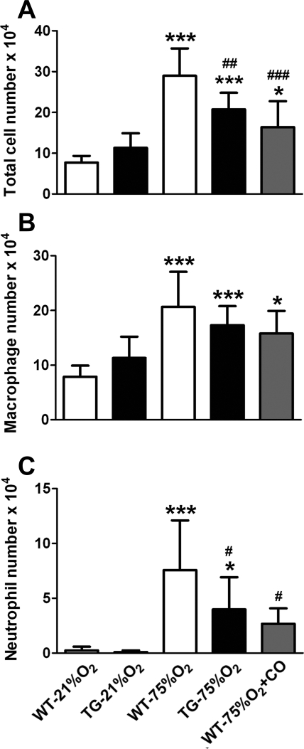
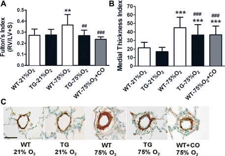
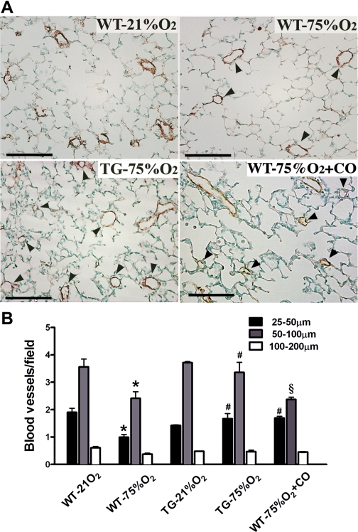
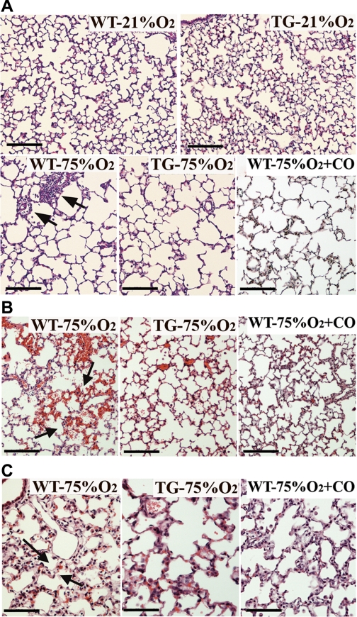
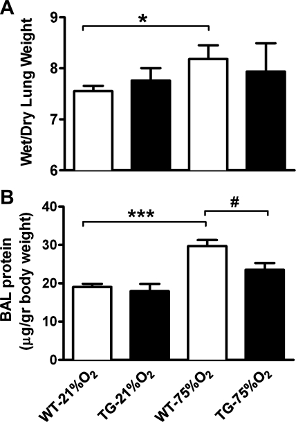
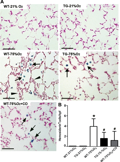
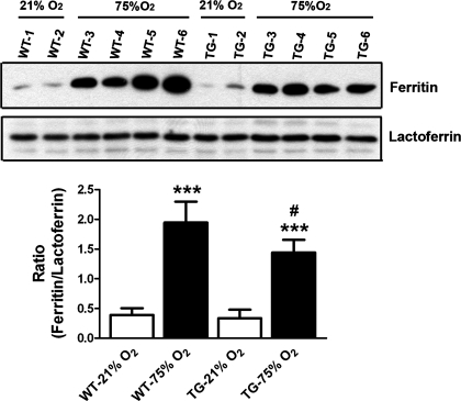
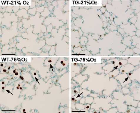
Bronchopulmonary dysplasia (BPD) is characterized by simplified alveolarization and arrested vascular development of the lung with associated evidence of endothelial dysfunction, inflammation, increased oxidative damage, and iron deposition. Heme oxygenase-1 (HO-1) has been reported to be protective in the pathogenesis of diseases of inflammatory and oxidative etiology. Because HO-1 is involved in the response to oxidative stress produced by hyperoxia and is critical for cellular heme and iron homeostasis, it could play a protective role in BPD. Therefore, we investigated the effect of HO-1 in hyperoxia-induced lung injury using a neonatal transgenic mouse model with constitutive lung-specific HO-1 overexpression. Hyperoxia triggered an increase in pulmonary inflammation, arterial remodeling, and right ventricular hypertrophy that was attenuated by HO-1 overexpression. In addition, hyperoxia led to pulmonary edema, hemosiderosis, and a decrease in blood vessel number, all of which were markedly improved in HO-1 overexpressing mice. The protective vascular response may be mediated at least in part by carbon monoxide, due to its anti-inflammatory, antiproliferative, and antiapoptotic properties. HO-1 overexpression, however, did not prevent alveolar simplification nor altered the levels of ferritin and lactoferrin, proteins involved in iron binding and transport. Thus the protective mechanisms elicited by HO-1 overexpression primarily preserve vascular growth and barrier function through iron-independent, antioxidant, and anti-inflammatory pathways.


Figures

Fig. 1.
Fig. 1.
Expression of heme oxygenase-1 (HO-1) in transgenic mice is not affected by hyperoxic exposure. A: mRNA levels of human HO-1 were assessed through quantitative PCR in total lung at 1 and 2 wk (w) following hyperoxia. Values are shown relative to normoxia. B: Western blot analysis of HO-1 protein in whole lungs from neonatal wild-type (WT) mice (21% O2) and transgenic (TG) mice after 2 wk of exposure to room air (21% O2) or hyperoxia (75%O2) after birth. Note that the antibody used detects both the constitutive human HO-1 and the endogenous murine HO-1. α-Tubulin was used as an internal control.
Fig. 2.
Fig. 2.
Hyperoxia-induced inflammatory cell influx in neonatal mouse lungs is attenuated by HO-1 overexpression and carbon monoxide (CO) inhalation. Total (A) and differential cell counts (B and C) were performed in bronchoalveolar lavage (BAL) fluid after 2 wk of exposure to room air (21% O2) or hyperoxia (75% O2) revealing a modest effect on the number of macrophages (B) and a significant decrease in the number of neutrophils (C) in hyperoxic HO-1 overexpressing mice and mice receiving inhaled CO, compared with WT controls (n = 10 mice per group). *P < 0.05, ***P < 0.001 vs. WT-21% O2; #P < 0.05, ##P < 0.01, ###P < 0.001 vs. WT-75% O2.
Fig. 3.
Fig. 3.
HO-1 overexpression and inhaled CO diminished the development of pulmonary vascular remodeling resulting from hyperoxia exposure. A: neonatal WT mice exposed for two wk to 75% O2 develop right ventricular hypertrophy (RVH) that is significantly reduced in HO-1 TG and CO-treated mice (250 ppm, 1 h daily; n = 15–18 mice per group). B: vascular remodeling of pulmonary arterioles was also partially but significantly decreased in TG and CO-treated mice compared with WT mice following 2 wk of hyperoxic exposure (n = 4–6 mice per group). C: Representative paraffin sections from lungs stained with α-smooth muscle actin for the visualization of medial wall thickness of pulmonary arterioles (25- to 75-μm diameter) shows a decreased smooth muscle layer thickening in HO-1 TG and CO receiving mice compared with WT mice following exposure to 75% O2. RV, right ventricle; LV, left ventricle; S, septum. **P < 0.01; ***P < 0.001 vs. WT-21% O2; ##P < 0.01, ###P < 0.001 vs. WT-75% O2. Scale bar in C = 30 μm.
Fig. 4.
Fig. 4.
Blood vessel loss after hyperoxic exposure is preserved by HO-1 overexpression and CO inhalation. A: lung sections stained for von Willebrand Factor (vWF), a marker of endothelial cells, revealed a greater loss of pulmonary blood vessels (arrowheads) per high-power field in lung sections from WT mice compared with HO-1 TG and CO-treated mice exposed to 75% O2. Scale bar = 100 μm. B: quantification of the number of small (25–50 μm), intermediate (50–100 μm), and large (100–200 μm) pulmonary vessels. Note that 75% O2 exposure in WT mice decreased the number of all three categories of vessels, while HO-1 overexpression preserved vessels of all sizes to numbers comparable to 21% O2-exposed mice (n = 3–4 mice per group) and CO treatment maintained the number of only the small vessels equivalent to those in normoxic mice. *P < 0.001 vs. WT-21% O2; #P < 0.001 vs. WT-75% O2; §P < 0.001 vs. TG −75% O2.
Fig. 5.
Fig. 5.
Lung histological examination showing the effects of hyperoxia, HO-1 overexpression and CO inhalation on alveolarization, hemorrhage, and edema. A: paraffin lung sections stained with hematoxylin and eosin demonstrated increased alveolar size and reduced septation in WT, HO-1 TG, and CO-treated mice exposed to hyperoxia (75%O2) 2 wk after birth, compared with normoxic (21% O2) mice. WT lungs also exhibited abundant perivascular mononuclear infiltration (arrows). B: areas of hemorrhage (seen as mild to moderate accumulation of red blood cells in the pulmonary parenchyma) as well as thickened alveolar walls, indicative of pulmonary edema (C, arrows) were abundant in hematoxylin and eosin-stained lung sections from WT mice exposed to 75% O2 but less common in lung sections from similarly exposed HO-1 TG and CO-treated mice (n = 4–6 mice per group). Scale bars in A and B = 100 μm and in C = 50 μm.
Fig. 6.
Fig. 6.
HO-1 TG mice exhibit decreased pulmonary edema and have lower BAL fluid protein content following hyperoxia. A: weight ratio of the wet-to-dry lung (n = 5–7) was significantly increased in WT neonatal mice 2 wk following 75% O2 exposure but not in HO-1 TG mice. B: total protein concentration in BAL fluid (n = 6–9) significantly increased in the WT hyperoxic group but not in the hyperoxic TG group compared with normoxic mice. *P < 0.05, ***P < 0.001 vs. WT-21% O2; #P < 0.05 vs. WT-75% O2.
Fig. 7.
Fig. 7.
Hemosiderosis induced by hyperoxia exposure is attenuated by HO-1 overexpression and CO inhalation. A: lung sections from WT, TG, and CO-treated mice stained with Perls Prussian blue iron revealed areas of severe hemosiderosis (arrows) in proximity to thickened alveolar walls (arrowheads) in WT mice exposed to 75% O2. Only moderate hemosiderosis was observed in hyperoxic HO-1 transgenic and CO-treated mice. B: hemosiderosis was quantified by counting iron-laden cells per high-power field in lung sections from each experimental group (n = 8). *P < 0.05 vs. WT- 21% O2; #P < 0.05 vs. WT-75% O2. Scale bar = 50 μm.
Fig. 8.
Fig. 8.
Induction of ferritin by heme-released iron is increased in hyperoxic WT mice. Representative Western blot of ferritin and lactoferrin proteins in whole lungs of neonatal mice from WT and HO-1 TG groups following 2 wk of room air (21% O2) or hyperoxia (75%O2) after birth. Quantification of the bands by densitometry revealed a significant increase in the ratio of ferritin to lactoferrin levels in WT hyperoxic mice compared with normoxia or to the TG group. Forty- to fifty-microliter aliquots of lung homogenates were electrophoresed and immunoblotted with anti-ferritin or anti-lactoferrin (see materials and methods). ***P < 0.001 vs. WT-21% O2; #P < 0.05 vs. WT-75% O2.
Fig. 9.
Fig. 9.
Hyperoxia-induced HO-1 expression is mainly restricted to pulmonary alveolar macrophages. Immunohistochemistry of paraffin lung sections with an antibody against murine HO-1 demonstrated minimal staining in mice that were exposed to room air (21% O2), whereas immunostaining was intense in macrophages (arrows) of both genotypes exposed to hyperoxia (75% O2) with significantly fewer stained macrophages in the hyperoxic HO-1 TG lungs, compared with WT hyperoxia. The evident epithelial HO-1 signal in the TG lung was due to cross-reactivity with the human HO-1 transgene expressed in type II epithelial cells. Scale bar = 50 μm.

References

    1. Abman SH. Impaired vascular endothelial growth factor signaling in the pathogenesis of neonatal pulmonary vascular disease. Adv Exp Med Biol 661: 323– 335, 2010 - PubMed
    1. Abman SH, Mourani PM, Sontag M. Bronchopulmonary dysplasia: a genetic disease. Pediatrics 122: 658– 659, 2008 - PubMed
    1. Abraham NG, Lavrovsky Y, Schwartzman ML, Stoltz RA, Levere RD, Gerritsen ME, Shibahara S, Kappas A. Transfection of the human heme oxygenase gene into rabbit coronary microvessel endothelial cells: protective effect against heme and hemoglobin toxicity. Proc Natl Acad Sci USA 92: 6798– 6802, 1995 - PMC - PubMed
    1. Actor JK, Hwang SA, Kruzel ML. Lactoferrin as a natural immune modulator. Curr Pharm Des 15: 1956– 1973, 2009 - PMC - PubMed
    1. Aslam M, Baveja R, Liang OD, Fernandez-Gonzalez A, Lee C, Mitsialis SA, Kourembanas S. Bone marrow stromal cells attenuate lung injury in a murine model of neonatal chronic lung disease. Am J Respir Crit Care Med 180: 1122– 1130, 2009 - PMC - PubMed

Publication types

LinkOut - more resources