. 2012:2012:601302.
doi: 10.1155/2012/601302. Epub 2012 Jan 12.

Optimization of human corneal endothelial cells for culture: the removal of corneal stromal fibroblast contamination using magnetic cell separation

Affiliations

Optimization of human corneal endothelial cells for culture: the removal of corneal stromal fibroblast contamination using magnetic cell separation

Gary S L Peh et al. Int J Biomater. 2012.

Abstract

The culture of human corneal endothelial cells (CECs) is critical for the development of suitable graft alternative on biodegradable material, specifically for endothelial keratoplasty, which can potentially alleviate the global shortage of transplant-grade donor corneas available. However, the propagation of slow proliferative CECs in vitro can be hindered by rapid growing stromal corneal fibroblasts (CSFs) that may be coisolated in some cases. The purpose of this study was to evaluate a strategy using magnetic cell separation (MACS) technique to deplete the contaminating CSFs from CEC cultures using antifibroblast magnetic microbeads. Separated "labeled" and "flow-through" cell fractions were collected separately, cultured, and morphologically assessed. Cells from the "flow-through" fraction displayed compact polygonal morphology and expressed Na(+)/K(+)ATPase indicative of corneal endothelial cells, whilst cells from the "labeled" fraction were mostly elongated and fibroblastic. A separation efficacy of 96.88% was observed. Hence, MACS technique can be useful in the depletion of contaminating CSFs from within a culture of CECs.


Figures
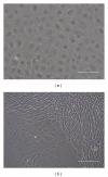
Figure 1
Figure 1
(a) Representative micrograph of a confluent homogeneous monolayer of successfully isolated human corneal endothelial cells at Day 14. (b) Representative micrograph of a failed isolation attempt resulting in stromal contamination at Day 7. The cellular boundary between the polygonal corneal endothelial cells (*) and the confluent elongated corneal stromal fibroblasts () can be clearly defined (scale bars = 100 μm).
Figure 2
Figure 2
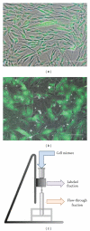
(a) Composite phase contrast and fluorescent picture of corneal stromal fibroblast labeled experimentally with CMFDA CellTracker Green dye. (b) Culture of CMFDA labeled corneal stromal fibroblast with human corneal endothelial cells 24 hours after mixing at a 1 : 1 ratio. (c) Schematic of the MACS setup used in the study (scale bar = 100 μm).
Figure 3
Figure 3
Following MACS of experimentally mixed human corneal endothelial cells and corneal stromal fibroblast using antifibroblast magnetic microbeads, cell fractions were subcultured as (a) the antifibroblast magnetic microbeads “labeled” fraction and (b) unlabeled “flow-through” fraction, 3 days after sorting. (Scale bar = 100 μm.) (c) A frequency histogram depicting the circularity of randomly selected cells in the “labeled” and the “flow-through” fractions. At least 100 cells from each fraction were counted.
Figure 4
Figure 4
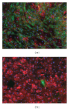
Composite fluorescent micrograph images of CMFDA (green) and Na+/K+ATPase pumps (red) of cultured cells established from (a) the “labeled” fraction and (b) the “flow-through” fraction. Cell nuclei were counterstained with DAPI in blue. (Scale bar = 100 μm.)

References

    1. Maurice DM. The location of the fluid pump in the cornea. Journal of Physiology. 1972;221(1):43–54. - PMC - PubMed
    1. Fischbarg J, Lim JJ. Role of cations, anions and carbonic anhydrase in fluid transport across rabbit corneal endothelium. Journal of Physiology. 1974;241(3):647–675. - PMC - PubMed
    1. Bourne WM. Clinical estimation of corneal endothelial pump function. Transactions of the American Ophthalmological Society. 1998;96:229–242. - PMC - PubMed
    1. Carlson KH, Bourne WM, McLaren JW, Brubaker RF. Variations in human corneal endothelial cell morphology and permeability to fluorescein with age. Experimental Eye Research. 1988;47(1):27–41. - PubMed
    1. Bourne WM, Nelson LIL, Hodge DO. Central corneal endothelial cell changes over a ten-year period. Investigative Ophthalmology and Visual Science. 1997;38(3):779–782. - PubMed

LinkOut - more resources