. 2011 Jan 1;7(3):213-222.
doi: 10.12989/sss.2011.7.3.213.

Biomimetics of the Extracellular Matrix: An Integrated Three-Dimensional Fiber-Hydrogel Composite for Cartilage Tissue Engineering

Affiliations

Biomimetics of the Extracellular Matrix: An Integrated Three-Dimensional Fiber-Hydrogel Composite for Cartilage Tissue Engineering

Jeannine Coburn et al. Smart Struct Syst. .

Abstract

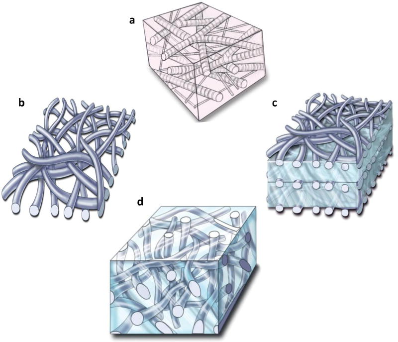
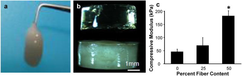
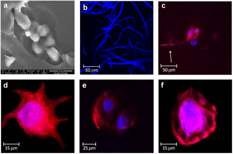
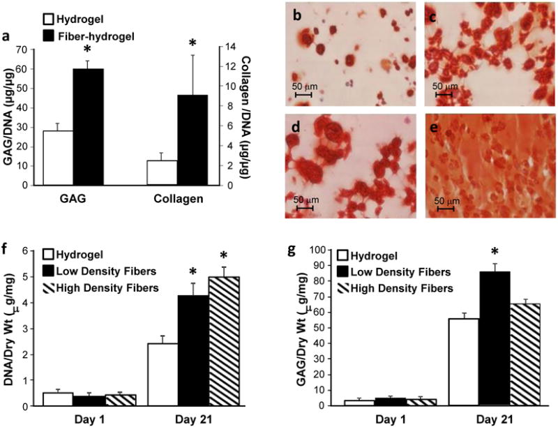
The native extracellular matrix (ECM) consists of an integrated fibrous protein network and proteoglycan-based ground (hydrogel) substance. We designed a novel electrospinning technique to engineer a three dimensional fiber-hydrogel composite that mimics the native ECM structure, is injectable, and has practical macroscale dimensions for clinically relevant tissue defects. In a model system of articular cartilage tissue engineering, the fiber-hydrogel composites enhanced the biological response of adult stem cells, with dynamic mechanical stimulation resulting in near native levels of extracellular matrix. This technology platform was expanded through structural and biochemical modification of the fibers including hydrophilic fibers containing chondroitin sulfate, a significant component of endogenous tissues, and hydrophobic fibers containing ECM microparticles.


Figures

Figure 1
Figure 1. Architectural framework of a native extracellular matrix (ECM) and biomimetic fiber-hydrogel scaffolds
a, The native ECM is composed of a fibrous protein network encapsulating a proteoglycan ground substance. b, Current electrospinning technologies produce a dense mat of fibers. c, The dense mats of fibers can be stacked with hydrogels to create a more three dimensional biomaterial scaffold. d, Novel electrospinning techniques allow synthesis of three-dimensional, low density fibers that can be combined with hydrogels to mimic the native ECM.
Figure 2
Figure 2. Fiber-hydrogel handling and physical properties
a, Hydrogel macromer solutions can be easily combined with the low density fibers and the resulting three-dimensional fiber-hydrogel composite is injectable before in situ hydrogel polymerization. b, After polymerization, the fibers are visible and homogenously distributed throughout the hydrogel (bottom), compared to the hydrogel alone (top). c, Incorporation of fibers into a hydrogel significantly enhanced compressive strength in a dose-dependent manner. *P≤0.05
Figure 3
Figure 3. Fiber-hydrogel ultrastructure and cellular morphology
a, Scanning electron microscopy focuses on fibers embedded in the hydrogel. b, Fluorescent staining and confocal microscopy of the fibers highlight the large pore size of the fiber network that creates a three dimensional structure that mimics the native ECM. c-e, Mesenchymal stem cell morphology in the mimetic fiber-hydrogel composites varies to include cells with multiple and long extensions and dividing cells with a round morphology. f, Cells in the hydrogel alone scaffolds have a round morphology.
Figure 4
Figure 4. Cartilage tissue development in the fiber-hydrogel scaffolds and response to mechanical stimuli
a, MSCs encapsulated in the lamellar fiber-hydrogel scaffolds and cultured in chondrogenic conditions produced significantly greater amounts of extracellular matrix. b, Safranin-O staining confirmed that MSCs secreted more proteoglycans in the fiber-hydrogel composites compared to the c, hydrogel alone scaffolds. d, Dynamic mechanical stimulation (+MS) produced a significantly greater cellular response with increased ECM deposition and homogenous distribution in the fiber-hydrogel composites as visualized by Safranin-O. e, compared to cells in the hydrogel alone. f, MSCs incorporated in the three dimensional fiber-hydrogel composites increased proliferation as the density of fibers increased. g, Proteoglycan secretion, a marker for cartilage formation, was greatest when MSCs were incorporated in fiber-hydrogel composites with a low density of fibers (low and high fiber density: 10% and 40% of dry weight, respectively). *P≤0.05
Figure 5
Figure 5. Chemical and biological flexibility of fibers for scaffolding
a, Three dimensional nanofibers were generated from a combination of PVA and chondroitin sulfate (CS). b, Histological staining with Safranin-O confirms the presence of the proteoglycan in the fibers. c, Segments of cartilage ECM were encapsulated in the fibers as visualized by scanning electron microscopy and d, Safranin-O staining for proteoglycans.

References

    1. Awad HA, Wickham MQ, Leddy HA, Gimble JM, Guilak F. Chondrogenic differentiation of adipose-derived adult stem cells in agarose, alginate, and gelatin scaffolds. Biomaterials. 2004;25(16):3211–3222. - PubMed
    1. Chiara G, Ranieri C. Cartilage and Bone Extracellular Matrix. Curr Pharm Des. 2009;15:1334–1348. - PubMed
    1. Engler AJ, Sen S, Sweeney HL, Discher DE. Matrix Elasticity Directs Stem Cell Lineage Specification. Cell. 2006;126(4):677–689. - PubMed
    1. Farndale RW, Buttle DJ, Barrett AJ. Improved quantitation and discrimination of sulphated glycosaminoglycans by use of dimethylmethylene blue. Biochim Biophys Acta (BBA) Gen Sub. 1986;883(2):173–177. - PubMed
    1. Gong JP, Katsuyama Y, Kurokawa T, Osada Y. Double-Network Hydrogels with Extremely High Mechanical Strength. Adv Mater. 2003;15(14):1155–1158.

LinkOut - more resources