. 2012 Feb;35(3):353-65.
doi: 10.1111/j.1460-9568.2011.07958.x.

Cysteine- and glycine-rich protein 1a is involved in spinal cord regeneration in adult zebrafish

Affiliations

Cysteine- and glycine-rich protein 1a is involved in spinal cord regeneration in adult zebrafish

Liping Ma et al. Eur J Neurosci. 2012 Feb.

Abstract

In contrast to mammals, adult zebrafish have the ability to regrow descending axons and gain locomotor recovery after spinal cord injury (SCI). In zebrafish, a decisive factor for successful spinal cord regeneration is the inherent ability of some neurons to regrow their axons via (re)expressing growth-associated genes during the regeneration period. The nucleus of the medial longitudinal fascicle (NMLF) is one of the nuclei capable of regenerative response after SCI. Using microarray analysis with laser capture microdissected NMLF, we show that cysteine- and glycine-rich protein (CRP)1a (encoded by the csrp1a gene in zebrafish), the function of which is largely unknown in the nervous system, was upregulated after SCI. In situ hybridization confirmed the upregulation of csrp1a expression in neurons during the axon growth phase after SCI, not only in the NMLF, but also in other nuclei capable of regeneration, such as the intermediate reticular formation and superior reticular formation. The upregulation of csrp1a expression in regenerating nuclei started at 3 days after SCI and continued to 21 days post-injury, the longest time point studied. In vivo knockdown of CRP1a expression using two different antisense morpholino oligonucleotides impaired axon regeneration and locomotor recovery when compared with a control morpholino, demonstrating that CRP1a upregulation is an important part of the innate regeneration capability in injured neurons of adult zebrafish. This study is the first to demonstrate the requirement of CRP1a for zebrafish spinal cord regeneration.


Figures

Figure 1
Figure 1. A cartoon diagramming the work flow for microarray analysis
Spinal cord injury (SCI) was performed with a complete transection at one millimeter caudal to the brainstem-spinal cord junction. Brains were removed at different time points after SCI and sections were collected. After staining with cresyl violet, laser capture microdissection was used to isolate the NMLF. Total RNA was extracted from the pooled NMLF tissue and was amplified. cDNA from amplified RNA was used for microarray analysis. Scale bar, 1 mm.
Figure 2
Figure 2. Csrp1a is upregulated in the NMLF at 11 days after SCI
(A) Graph of normalized fold changes for GAP-43 and csrp1a from microarray analysis. (B) Quantitative real-time PCR (qPCR) shows that both GAP-43 and csrp1a are upregulated in the NMLF at 11 days after SCI. (C) qPCR shows that csrp1a levels are slightly downregulated in the caudal part of the spinal cord at 11 days after SCI. n = 3 experiments. * P < 0.05, t-test; Mean values ± SEM are shown.
Figure 3
Figure 3. Csrp1a is upregulated in the NMLF, IMRF and SRF at 11 days after SCI
(A) In situ hybridization was performed to study the expression of csrp1a in regenerative nuclei. Representative images depict csrp1a positive neurons in NMLF, IMRF and SRF at 11 days after SCI. Schematic drawings of the NMLF, IMRF and SRF are shown. Positive signal for csrp1a is observed in neurons, i.e. cells with a diameter more than 13 µm. More positive neurons for csrp1a were observed in all three nuclei after SCI when compared with sham-lesioned control. With sense probe, no significant signal was observed. Arrows in the schematic drawings indicate the corresponding nucleus. Arrows in images indicate the brain midline. (B) Quantification shows that csrp1a is upregulated in NMLF, IMRF and SRF at 11 days after SCI. NMLF (nucleus of the medial longitudinal fascicle), n = 6 fish; IMRF (intermediate reticular formation), n = 3 fish; SRF (superior reticular formation), n = 3 fish. * P < 0.05, t-test; Mean values ± SEM are shown. Scale bar, 50 µm.
Figure 4
Figure 4. Csrp1a is upregulated in NeuN-positive neurons in regenerative nuclei after SCI
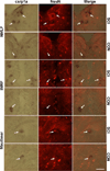
Double staining of csrp1a (in situ hybridization) and NeuN (immunohistochemistry) was performed to examine whether csrp1a-positive cells are neurons or not. The signal for NeuN locates in nucleus and csrp1a locates in cytoplasm. Cells positive for csrp1a are also labeled by NeuN, demonstrating that csrp1a is expressed by neurons. More double labeling cells are observed at 11 days after SCI in regenerative nuclei, such as NMLF (upper two rows) and IMRF (middle two rows), when compared to control, while no significant upregulation of csrp1a expression is found after SCI in Mauthner cells (lower two rows) which are not capable to regenerate after SCI. n = 3 experiments. Scale bar, 50 µm.
Figure 5
Figure 5. Csrp1a is upregulated in the axon regeneration phase after SCI
In situ hybridization was performed to investigate the regulation pattern of csrp1a expression after SCI. Three different time points were included, 3 days, 11 days and 21 days after SCI. Quantification shows that csrp1a is slightly upregulated at 3 days. The expression of csrp1a is highly induced at 11 days post-injury and this upregulation continues to 21 days, the longest time point tested. Similar regulation pattern of csrp1a expression are observed in NMLF (A), IMRF (B) and SRF (C). n = 3 fish for each time points. * P < 0.05, two-way ANOVA with Tukey post-hoc test; Mean values ± SEM are shown.
Figure 6
Figure 6. Application of CRP1 MO inhibits locomotor recovery after SCI
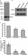
(A) The CRP1 antibody used for Western blot analysis only detects one band with correct size for CRP1, demonstrating the specificity of the antibody. Signal for CRP1 was detected with both samples from zebrafish spinal cord and brain. SC: spinal cord. (B–C) Both CRP1 MO1 and CRP1 MO2 knock down CRP1 expression. Two millimeter spinal cord tissue, centered on the transection site, was collected at 11 days after SCI and MO treatment. α-tubulin serves as a loading control. CRP1 MO1 and CRP1 MO2 exhibited 30% and 40% knock-down effect on CRP1 expression compared to control MO, respectively, as demonstrated by the Western blot analysis (B) and the densitometric analysis (C). (D)Total distance moved by animals treated with control (CON) MO, or CRP1 MO1, or CRP1 MO2 was measured during 5 minutes trial periods by video recording 6 weeks after MO application. CRP1 MO1 (n = 12 fish) or CRP1 MO2 (n = 5 fish) treatments reduce the total distance moved when compared to CON MO treatment (n = 9 fish). A–C, n = 3 experiments. * P < 0.05, one-way ANOVA with Tukey post-hoc test; Mean values ± SEM are shown.
Figure 7
Figure 7. CRP1 MO inhibits axon regeneration after SCI
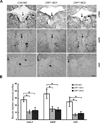
(A) Representative images of retrogradely labeled neurons in NMLF, IMRF and SRF. The number of retrogradely labeled neurons in the NMLF, IMRF and SRF (biocytin label) is measured 6 weeks after SCI and MO application. Biocytin was applied 4 mm caudal to the lesion site and was detected 24 hours later. CRP1 MO1 and CRP1 MO2 treatments reduce biocytin-labeled neurons in all nuclei studied when compared to CON MO. Arrows in images indicate the brain midline. (B) Quantification shows a reduction in number of biocytin-labeled neurons in fish received CRP1 MO1 or CRP1 MO2 compared to fish received CON MO. * P < 0.05, one-way ANOVA with Tukey post-hoc test; Mean values ± SEM are shown. Scale bar, 50 µm.
Figure 8
Figure 8. CRP1 MO1 does not affect cell viability
(A) Representative images of neurons with fluorescein signal in the NMLF 6 weeks after MO application. The standard control MO and CRP1 MO1 were tagged with fluorescein and this signal is still detectable at 6 weeks after MO treatments. No difference was found between the numbers of cells positive for fluorescein in animals treated with CON MO or CRP1 MO1. Arrows in images indicate the brain midline. (B) Quantification shows no difference of fluorescein positive cells in the NMLF in animals treated with CON MO (n = 3 fish) or CRP1 MO1 (n = 4 fish). * P < 0.05, t-test; Mean values ± SEM are shown. Scale bar, 50 µm.

References

    1. Becker CG, Becker T. Adult zebrafish as a model for successful central nervous system regeneration. Restor Neurol Neurosci. 2008;26:71–80. - PubMed
    1. Becker CG, Lieberoth BC, Morellini F, Feldner J, Becker T, Schachner M. L1.1 is involved in spinal cord regeneration in adult zebrafish. J Neurosci. 2004;24:7837–7842. - PMC - PubMed
    1. Becker T, Bernhardt RR, Reinhard E, Wullimann MF, Tongiorgi E, Schachner M. Readiness of zebrafish brain neurons to regenerate a spinal axon correlates with differential expression of specific cell recognition molecules. J Neurosci. 1998;18:5789–5803. - PMC - PubMed
    1. Becker T, Wullimann MF, Becker CG, Bernhardt RR, Schachner M. Axonal regrowth after spinal cord transection in adult zebrafish. J Comp Neurol. 1997;377:577–595. - PubMed
    1. Bernhardt RR. Cellular and molecular bases of axonal regeneration in the fish central nervous system. Exp Neurol. 1999;157:223–240. - PubMed

Publication types

MeSH terms