

Multifunctional CD4⁺ T cells in patients with American cutaneous leishmaniasis
- PMID: 22288594
- PMCID: PMC3374283
- DOI: 10.1111/j.1365-2249.2011.04536.x
Multifunctional CD4⁺ T cells in patients with American cutaneous leishmaniasis
Abstract
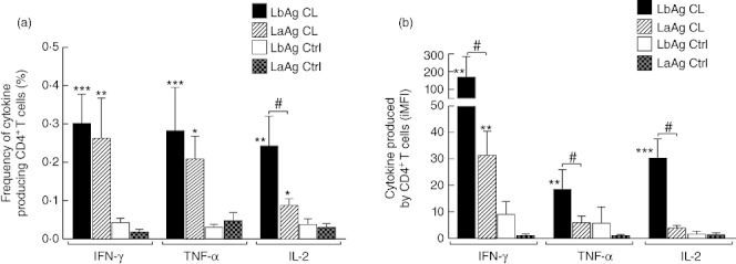
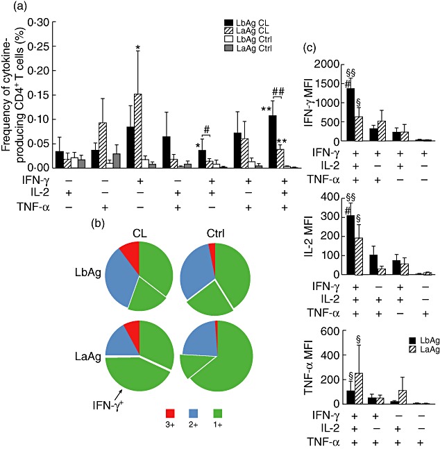
Leishmaniasis is a group of important parasitic diseases affecting millions worldwide. To understand more clearly the quality of T helper type 1 (Th1) response stimulated after Leishmania infection, we applied a multiparametric flow cytometry protocol to evaluate multifunctional T cells induced by crude antigen extracts obtained from promastigotes of Leishmania braziliensis (LbAg) and Leishmania amazonensis (LaAg) in peripheral blood mononuclear cells from healed cutaneous leishmaniasis patients. Although no significant difference was detected in the percentage of total interferon (IFN)-γ-producing CD4(+) T cells induced by both antigens, multiparametric flow cytometry analysis revealed clear differences in the quality of Th1 responses. LbAg induced an important proportion of multifunctional CD4(+) T cells (28% of the total Th1 response evaluated), whereas LaAg induced predominantly single-positive cells (68%), and 57% of those were IFN-γ single-positives. Multifunctional CD4(+) T cells showed the highest mean fluorescence intensity (MFI) for the three Th1 cytokines assessed and MFIs for IFN-γ and interleukin-2 from those cells stimulated with LbAg were significantly higher than those obtained after LaAg stimulation. These major differences observed in the generation of multifunctional CD4(+) T cells suggest that the quality of the Th1 response induced by L. amazonensis antigens can be involved in the mechanisms responsible for the high susceptibility observed in L. amazonensis-infected individuals. Ultimately, our results call attention to the importance of studying a Th1 response regarding its quality, not just its magnitude, and indicate that this kind of evaluation might help understanding of the complex and diverse immunopathogenesis of American tegumentary leishmaniasis.
© 2011 The Authors. Clinical and Experimental Immunology © 2011 British Society for Immunology.
Figures
Similar articles
-
Flow cytometric determination of cellular sources and frequencies of key cytokine-producing lymphocytes directed against recombinant LACK and soluble Leishmania antigen in human cutaneous leishmaniasis.Infect Immun. 2001 May;69(5):3232-9. doi: 10.1128/IAI.69.5.3232-3239.2001. Infect Immun. 2001. PMID: 11292745 Free PMC article.
-
T-cell responsiveness of American cutaneous leishmaniasis patients to purified Leishmania pifanoi amastigote antigens and Leishmania braziliensis promastigote antigens: immunologic patterns associated with cure.Exp Parasitol. 1996 Nov;84(2):144-55. doi: 10.1006/expr.1996.0100. Exp Parasitol. 1996. PMID: 8932764
-
Contribution of Leishmania braziliensis antigen-specific CD4+ T, CD8+ T, NK and CD3+CD56+NKT cells in the immunopathogenesis of cutaneous leishmaniasis patients: Cytotoxic, activation and exhaustion profiles.PLoS One. 2020 Mar 23;15(3):e0229400. doi: 10.1371/journal.pone.0229400. eCollection 2020. PLoS One. 2020. PMID: 32203546 Free PMC article.
-
Immunopathogenic competences of Leishmania (V.) braziliensis and L. (L.) amazonensis in American cutaneous leishmaniasis.Parasite Immunol. 2009 Aug;31(8):423-31. doi: 10.1111/j.1365-3024.2009.01116.x. Parasite Immunol. 2009. PMID: 19646206 Review.
-
[Immunopathology of American tegumentary leishmaniasis].Acta Cient Venez. 1998;49(1):42-56. Acta Cient Venez. 1998. PMID: 10205916 Review. Spanish.
Cited by
-
Design of multi-epitope peptides containing HLA class-I and class-II-restricted epitopes derived from immunogenic Leishmania proteins, and evaluation of CD4+ and CD8+ T cell responses induced in cured cutaneous leishmaniasis subjects.PLoS Negl Trop Dis. 2020 Mar 16;14(3):e0008093. doi: 10.1371/journal.pntd.0008093. eCollection 2020 Mar. PLoS Negl Trop Dis. 2020. PMID: 32176691 Free PMC article.
-
Insulin-like growth factor-I induces arginase activity in Leishmania amazonensis amastigote-infected macrophages through a cytokine-independent mechanism.Mediators Inflamm. 2014;2014:475919. doi: 10.1155/2014/475919. Epub 2014 Sep 9. Mediators Inflamm. 2014. PMID: 25294956 Free PMC article.
-
Immunopathogenesis of non-healing American cutaneous leishmaniasis and progressive visceral leishmaniasis.Semin Immunopathol. 2012 Nov;34(6):735-51. doi: 10.1007/s00281-012-0350-8. Epub 2012 Oct 11. Semin Immunopathol. 2012. PMID: 23053396 Free PMC article. Review.
-
Attenuated and replication-competent vaccinia virus strains M65 and M101 with distinct biology and immunogenicity as potential vaccine candidates against pathogens.J Virol. 2013 Jun;87(12):6955-74. doi: 10.1128/JVI.03013-12. Epub 2013 Apr 17. J Virol. 2013. PMID: 23596295 Free PMC article.
-
American tegumentary leishmaniasis: T-cell differentiation profile of cutaneous and mucosal forms-co-infection with Trypanosoma cruzi.Med Microbiol Immunol. 2016 Aug;205(4):353-69. doi: 10.1007/s00430-016-0455-0. Epub 2016 Apr 4. Med Microbiol Immunol. 2016. PMID: 27040974
References
-
- World Health Organization (WHO) Leishmaniasis. Available at: http://www.who.int/leishmaniasis/en/ (accessed 27 June 2011)
-
- Schwartz E, Hatz C, Blum J. New world cutaneous leishmaniasis in travellers. Lancet Infect Dis. 2006;6:342–9. - PubMed
-
- de Oliveira-Neto MP, Mattos MS, Perez MA, et al. American tegumentary leishmaniasis (ATL) in Rio de Janeiro State, Brazil: main clinical and epidemiologic characteristics. Int J Dermatol. 2000;39:506–14. - PubMed
Publication types
MeSH terms
Substances
LinkOut - more resources
Full Text Sources
Research Materials
Miscellaneous
