. 2012 Mar;167(3):543-55.
doi: 10.1111/j.1365-2249.2011.04540.x.

Stromal cells from term fetal membrane are highly suppressive in allogeneic settings in vitro

Affiliations

Stromal cells from term fetal membrane are highly suppressive in allogeneic settings in vitro

H Karlsson et al. Clin Exp Immunol. 2012 Mar.

Abstract

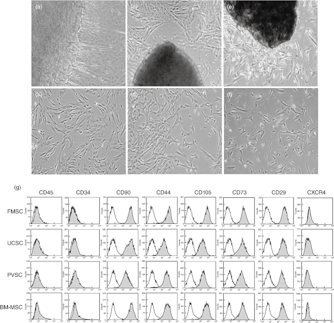
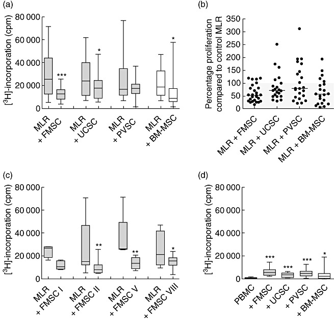
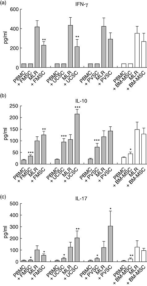
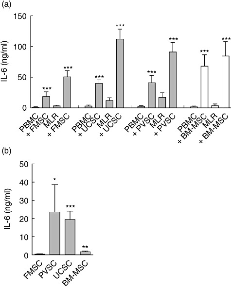
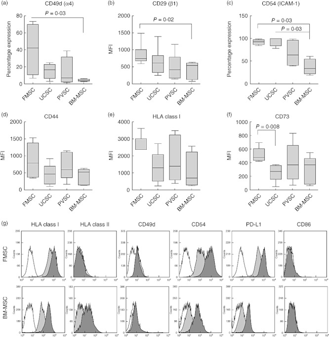
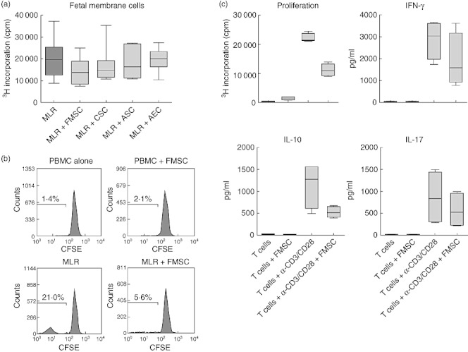
Bone marrow-derived mesenchymal stromal cells (BM-MSCs) have immunosuppressive properties and have been used to treat steroid-refractory acute graft-versus-host disease (GVHD) in stem cell transplant patients. Cells with similar capacities can also be found in term placental tissue. We have isolated stromal cells from term fetal membrane (FMSCs), umbilical cords (UCSCs) and placental villi (PVSCs) as well as from bone marrow and compared their immunoregulatory capacity in allogeneic settings. We found that FMSCs and UCSCs suppressed proliferation significantly in mixed lymphocyte reactions (MLRs), whereas PVSCs showed inconsistent suppressive effects. When added to MLR cultures, FMSCs suppressed the production of interferon (IFN)-γ and interleukin (IL)-17, whereas UCSCs and PVSCs promoted the production of IL-17 instead. Secretion of IL-10 was increased after addition of FMSCs and UCSCs. In this setting, BM-MSCs had no significant effect on secretion of IFN-γ, IL-17 or IL-10 in MLR cultures. When analysing the expression of adhesion markers, we noted that FMSCs expressed the highest levels of CD29 (β1), CD49d (α4) and CD54 (ICAM-1) compared to the other types of stromal cells. Thus, our data indicate that stromal cells isolated from term fetal membrane have great immunosuppressive capacity in terms of proliferation and production of proinflammatory cytokines from alloreactive T cells, and also promote anti-inflammatory IL-10. They express high levels of integrins that may be of importance in homing to inflamed tissues. Fetal membrane may provide a valuable source of cells with immunosuppressive properties and could possibly be used for treatment of acute GVHD and other inflammatory disorders.


Figures

Fig. 1
Fig. 1
Photomicrographs and phenotypic characterization of stromal cells from placental tissues. Panels (a), (c) and (e) illustrate primary cultures of fetal membrane, umbilical cord and placental villi, respectively, with cells protruding from the tissues. Original magnification: ×4. Panels (b), (d) and (f) show stromal cells from fetal membrane, umbilical cord and placental villi, respectively, at passage 3. Original magnification: ×10. (g) Phenotypic analysis of stromal cells isolated from fetal membrane (FMSC), umbilical cord (UCSC), placental villi (PVSC) and bone marrow (BM-MSC) in passage 3. Grey and white histograms depict the corresponding markers and isotype controls, respectively.
Fig. 2
Fig. 2
Fetal membrane stromal cells show strong immunosuppressive capacity. (a) Effect of proliferation after addition of stromal cells from fetal membrane (FMSCs) [n = 27 mixed lymphocyte reactions (MLRs) with different responder peripheral blood mononuclear cells (PBMCs)], umbilical cord (UCSCs) (n = 21 MLRs), placental villi (PVSCs) (n = 21 MLRs) and bone marrow-derived mesenchymal stem cells (BM-MSCs) (n = 21 MLRs) to MLR cultures (responder PBMC to stromal cell ratio 10:1). Proliferation was measured on day 6. (b) Percentage proliferation in MLRs after addition of the indicated type of stromal cells compared to control MLRs without stromal cells. (c) Alloantigen-induced proliferation with or without FMSCs from four different donors (from left to right: n = 4, 5, 8 and 8 responder PBMCs, respectively). (d) Stromal cell-induced proliferation of PBMCs (n = 15). The data are presented as box-and-whiskers with the maximum, minimum, median, 25th and 75th quartiles, and were generated by using FMSC, UCSC, PVSC and BM-MSC from four, three, three and three donors, respectively. *P < 0·05; **P < 0·01; ***P < 0·001 compared to the MLRs (panels a and c) or PBMCs (panel d) in the absence of stromal cells, as analysed by Wilcoxon's matched-pairs test.
Fig. 3
Fig. 3
Fetal membrane stromal cells suppress interferon (IFN)-γ and interleukin (IL)-17 production, but induce IL-10 production. Peripheral blood mononuclear cells (PBMCs) were stimulated with irradiated allogeneic PBMCs [mixed lymphocyte reactions (MLRs)] or were left unstimulated (PBMCs) in the presence or absence of stromal cells from fetal membrane (FMSCs) (n = 30 MLRs with different responder PBMCs), umbilical cord (UCSCs) (n = 22 MLRs), placental villi (PVSCs) (n = 22 MLRs) and bone marrow-derived mesenchymal stem cells (BM-MSCs) (n = 20 MLRs). Supernatants were harvested on day 5 and levels of (a) IFN-γ, (b) IL-17 and (c) IFN-γ were analysed by enzyme-linked immunosorbent assay. The data are presented as the mean cytokine production ± standard error of the mean, and were generated by using FMSC, UCSC, PVSC and BM-MSC from four, three, three and three donors, respectively. *P < 0·05; **P < 0·01; ***P < 0·001 compared to the MLRs or PBMCs in the absence of stromal cells, as analysed by Wilcoxon's matched-pairs test.
Fig. 4
Fig. 4
Fetal membrane stromal cells produce low levels of interleukin (IL)-6. (a) Peripheral blood mononuclear cells (PBMCs) were stimulated with irradiated allogeneic PBMCs [mixed lymphocyte reactions (MLRs)] or left unstimulated (PBMCs) in the presence or absence of stromal cells from fetal membrane (FMSCs) (n = 30 MLRs with different responder PBMCs), umbilical cord (UCSCs) (n = 22 MLRs), placental villi (PVSCs) (n = 22 MLRs) and bone marrow-derived mesenchymal stem cells (BM-MSCs) (n = 20 MLRs). Supernatants were harvested on day 5 and levels of IL-6 were determined by enzyme-linked immunosorbent assay. (b) Levels of IL-6 were determined in supernatants from the type of stromal cell indicated after 5 days of culture. The data are presented as the mean cytokine production ± standard error of the mean and were generated by using FMSC, UCSC, PVSC and BM-MSC from four, three, three and three donors, respectively. *P < 0·05; **P < 0·01; ***P < 0·001 compared to the MLRs or PBMCs in the absence of stromal cells, as analysed by Wilcoxon's matched-pairs test (a). The Mann–Whitney U-test was used to compare the levels of IL-6 produced from different types of stromal cells relative to the level produced by FMSCs (b).
Fig. 5
Fig. 5
Fetal membrane stromal cells express high levels of adhesion molecules. (a–f) Expression of different markers on stromal cells from fetal membrane (FMSCs), umbilical cord (UCSCs), placental villi (PVSCs) and bone marrow-derived mesenchymal stromal cells (BM-MSCs) were analysed by flow cytometry (n = 4 for each type of stromal cell). The Mann–Whitney U-test was used to compare the mean fluorescence intensity (MFI) or the percentage expression of cell surface markers on different types of stromal cells. The data are presented as box-and-whiskers with the maximum, minimum, median, 25th and 75th quartiles. (g) Representative histograms of expression of different markers on unstimulated (light grey histograms) or interferon (IFN)-γ-stimulated (dark grey histograms) FMSCs (n = 6) and BM-MSCs (n = 3). White histograms depict isotype controls.
Fig. 6
Fig. 6
Fetal membrane stromal cells mediate their suppressive effect on T cells. (a) Effect on proliferation in mixed lymphocyte reactions (MLRs) [n = 7 different responder peripheral blood mononuclear cells (PBMCs)] after addition of stromal cells generated from intact fetal membrane (FMSC), separated chorion (CSC) or amnion (ASC) or amnion epithelial cells (AEC) (n = 2 for each type of stromal cell). (b) Responder PBMCs were labelled with carboxyfluorescein succinimidyl ester (CFSE) before stimulation with allogeneic PBMC (MLR) and the effect of FMSCs were analysed on T cells by gating on CD3+ cells. (c) Effect of FMSCs on proliferation and cytokine production from purified CD3+ T cells after stimulation with anti-CD3- and CD28-coated beads (n = 4). The data are presented as box-and-whiskers with the maximum, minimum, median, 25th and 75th quartiles.

References

    1. Thomas ED, Buckner CD, Banaji M, et al. One hundred patients with acute leukemia treated by chemotherapy, total body irradiation, and allogeneic marrow transplantation. Blood. 1977;49:511–33. - PubMed
    1. Hoogerbrugge PM, Brouwer OF, Bordigoni P, et al. Allogeneic bone marrow transplantation for lysosomal storage diseases. The European Group for Bone Marrow Transplantation. Lancet. 1995;345:1398–402. - PubMed
    1. Le Blanc K, Tammik L, Sundberg B, Haynesworth SE, Ringden O. Mesenchymal stem cells inhibit and stimulate mixed lymphocyte cultures and mitogenic responses independently of the major histocompatibility complex. Scand J Immunol. 2003;57:11–20. - PubMed
    1. Tse WT, Pendleton JD, Beyer WM, Egalka MC, Guinan EC. Suppression of allogeneic T-cell proliferation by human marrow stromal cells: implications in transplantation. Transplantation. 2003;75:389–97. - PubMed
    1. Ren G, Zhang L, Zhao X, et al. Mesenchymal stem cell-mediated immunosuppression occurs via concerted action of chemokines and nitric oxide. Cell Stem Cell. 2008;2:141–50. - PubMed

Publication types

MeSH terms