. 2012 Jan 30:11:14.
doi: 10.1186/1475-2859-11-14.

One-step generation of error-prone PCR libraries using Gateway® technology

Affiliations

One-step generation of error-prone PCR libraries using Gateway® technology

Antoine Gruet et al. Microb Cell Fact. .

Abstract

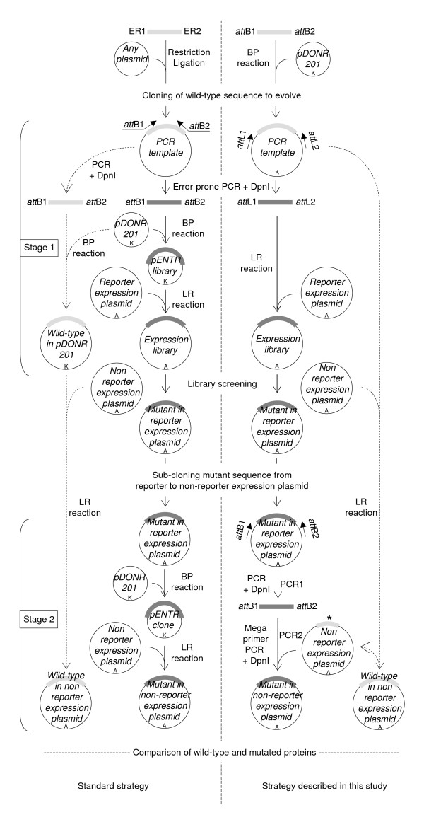
Background: Error-prone PCR (epPCR) libraries are one of the tools used in directed evolution. The Gateway® technology allows constructing epPCR libraries virtually devoid of any background (i.e., of insert-free plasmid), but requires two steps: the BP and the LR reactions and the associated E. coli cell transformations and plasmid purifications.

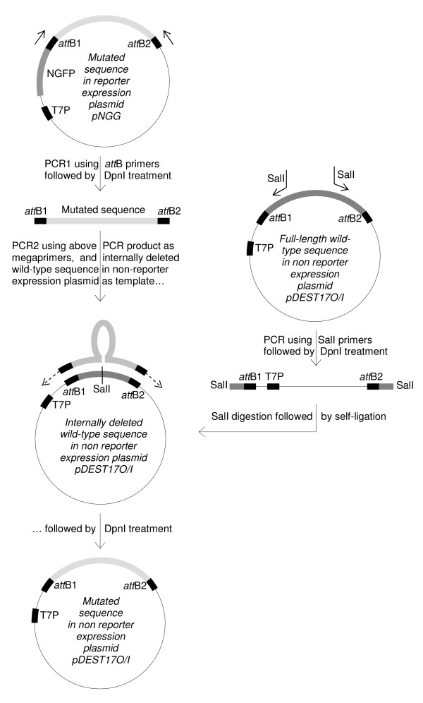
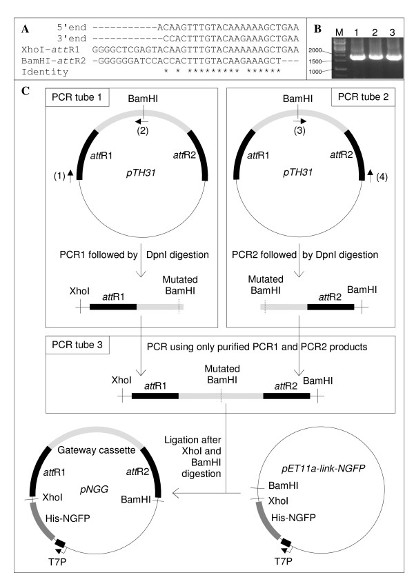
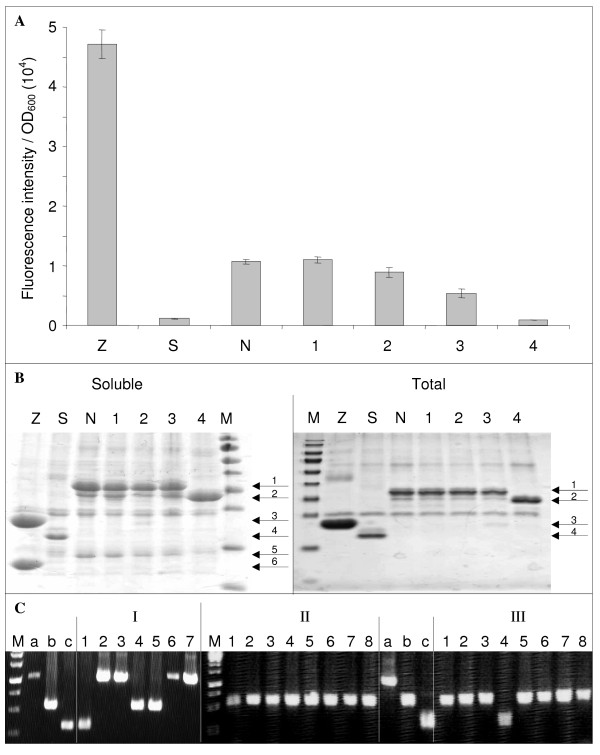
Results: We describe a method for making epPCR libraries in Gateway® plasmids using an LR reaction without intermediate BP reaction. We also describe a BP-free and LR-free sub-cloning method for in-frame transferring the coding sequence of selected clones from the plasmid used to screen the library to another one devoid of tag used for screening (such as the green fluorescent protein). We report preliminary results of a directed evolution program using this method.

Conclusions: The one-step method enables producing epPCR libraries of as high complexity and quality as does the regular, two-step, protocol for half the amount of work. In addition, it contributes to preserve the original complexity of the epPCR product.


Figures

Figure 1
Figure 1
Overview of the method. The left flowchart is the standard strategy and the right flowchart is the strategy described in this study. Brackets on the left indicate the two stages of the strategy: the epPCR library construction (stage1) and the sub-cloning of mutant inserts from reporter to non-reporter expression plasmid (stage 2). ER1 and ER2 denote restriction sites used to clone the sequence to evolve and create the PCR template of the standard strategy. Inner arrows with continuous lines are the core of the method. Outer arrows with dashed lines indicate the pathways used to transfer the wt sequence to the non-reporter expression plasmid, and (in the right flowchart only) to create the internally deleted wtNTAIL in pDEST17O/I. Mutated and wt sequences are represented by thick dark and light grey lines, respectively. The internally deleted wt sequence is denoted by an asterisk. Antibiotic resistance markers are indicated: A, ampicillin resistance; K, kanamycin resistance.
Figure 2
Figure 2
Schematic of the sub-cloning of a mutated sequence from reporter to non-reporter expression plasmid. From top to bottom, the DNA fragments amplified by PCR between the T7prom and attB2 primers are 1,046 bp, 333 bp, and 560 bp in length, respectively. On the right is illustrated how inverse PCR was used to internally delete 227 bp from the wtNTAIL sequence in pDEST17O/I. Plasmids are not at scale.
Figure 3
Figure 3
Construction of pNGG. (A) Alignment of (from top to bottom): the 5' and 3' ends of pTH31 Gateway® cassette; XhoI-attR1 PCR primer; BamHI-attR2 PCR primer. Sequence identity is denoted by asterisks below the alignment. (B) The Gateway® cassette was PCR amplified using pTH31 as template, and either primer XhoI-attR1 alone (lane 1), primer BamHI-attR2 alone (lane 2), or primers XhoI-attR1 and BamHI-attR2 (lane 3). Markers size is indicated on the left in base pairs. (C) "Two-halves" making of pNGG. The plasmids are not at scale. Light grey, Gateway cassette. Black, attR recombination sites. Primer (1), XhoI-attR1. Primer (2), BamHI-mut-R. Primer (3), BamHI-mut-F. Primer (4), BamHI-attR2 (Table 3).
Figure 4
Figure 4
Representative results of library screening and sub-cloning experiments. (A) The fluorescence to OD600 ratio (mean value and standard deviation of a triplicate experiment) of the clones indicated on the × axis were determined as described in Methods. Z, Leucine zippers; S, Stop-NTAIL; N, wtNTAIL; 1-3, full length mutated NTAIL; 4, truncated NTAIL mutant. (B) His-tagged proteins expressed by clones Z, S, N, 1-4 (Figure 4A) were purified by affinity chromatography on IMAC as described in Methods, and were analyzed by SDS-PAGE using 15% polyacrylamide gels and Coomassie blue staining. Soluble, His-tagged proteins were purified under non denaturing conditions from the soluble fraction of the E. coli lysate. Total, His-tagged proteins were purified under denaturing conditions from total E. coli lysate. Soluble and total fractions were obtained from a duplicate culture. M, molecular size markers (from top to bottom: 170, 130, 100, 70, 55, 40, 35, 25, 15, 10 kDa). Arrows indicate the different purified proteins: 1, NGFP- wtNTAIL and NGFP-full-length NTAIL variants (34 kDa); 2, NGFP-truncated NTAIL variant 4 (29.4 kDa); 3, NGP-Z (22.8 kDa); 4, NGFP (i.e., Stop-NTAIL) (20.4 kDa); 5, XD-CGFP (15.5 kDa); 6, Z-CGFP (13.3 kDa). (C) PCR screening of mutated NTAIL sub-cloning experiment from pNGG to pDEST17O/I. PCR control a, 1,046 bp; b, 560 bp; c, 333 bp. PCR screening II and III were run in the same gel along with controls a, b, and c. M, molecular size markers (from top to bottom: 2000, 1500, 1000, 750, 500, 250 bp).

References

    1. Vincentelli R, Bignon C, Gruez A, Canaan S, Sulzenbacher G, Tegoni M, Campanacci V, Cambillau C. Medium-scale structural genomics: strategies for protein expression and crystallization. Acc Chem Res. 2003;36(3):165–172. doi: 10.1021/ar010130s. - DOI - PubMed
    1. Stemmer WP. DNA shuffling by random fragmentation and reassembly: in vitro recombination for molecular evolution. Proc Natl Acad Sci USA. 1994;91(22):10747–10751. doi: 10.1073/pnas.91.22.10747. - DOI - PMC - PubMed
    1. Waldo GS, Standish BM, Berendzen J, Terwilliger TC. Rapid protein-folding assay using green fluorescent protein. Nat Biotechnol. 1999;17(7):691–695. doi: 10.1038/10904. - DOI - PubMed
    1. van den Berg S, Lofdahl PA, Hard T, Berglund H. Improved solubility of TEV protease by directed evolution. J Biotechnol. 2006;121(3):291–298. doi: 10.1016/j.jbiotec.2005.08.006. - DOI - PubMed
    1. Heddle C, Mazaleyrat SL. Development of a screening platform for directed evolution using the reef coral fluorescent protein ZsGreen as a solubility reporter. Protein Eng Des Sel. 2007;20(7):327–337. doi: 10.1093/protein/gzm024. - DOI - PubMed

Publication types

LinkOut - more resources