. 2012 Jan 30:11:5.
doi: 10.1186/1476-4598-11-5.

miR-31 and its host gene lncRNA LOC554202 are regulated by promoter hypermethylation in triple-negative breast cancer

Affiliations

miR-31 and its host gene lncRNA LOC554202 are regulated by promoter hypermethylation in triple-negative breast cancer

Katarzyna Augoff et al. Mol Cancer. .

Abstract

Background: microRNAs have been established as powerful regulators of gene expression in normal physiological as well as in pathological conditions, including cancer progression and metastasis. Recent studies have demonstrated a key role of miR-31 in the progression and metastasis of breast cancer. Downregulation of miR-31 enhances several steps of the invasion-metastasis cascade in breast cancer, i.e., local invasion, extravasation and survival in the circulation system, and metastatic colonization of distant sites. miR-31 exerts its metastasis-suppressor activity by targeting a cohort of pro-metastatic genes, including RhoA and WAVE3. The molecular mechanisms that lead to the loss of miR-31 and the activation of its pro-metastatic target genes during these specific steps of the invasion-metastasis cascade are however unknown.

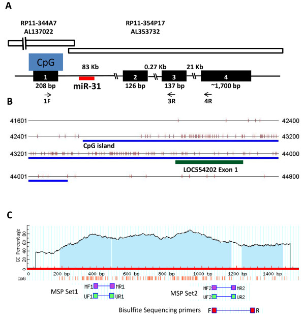
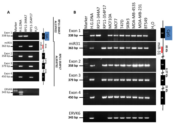
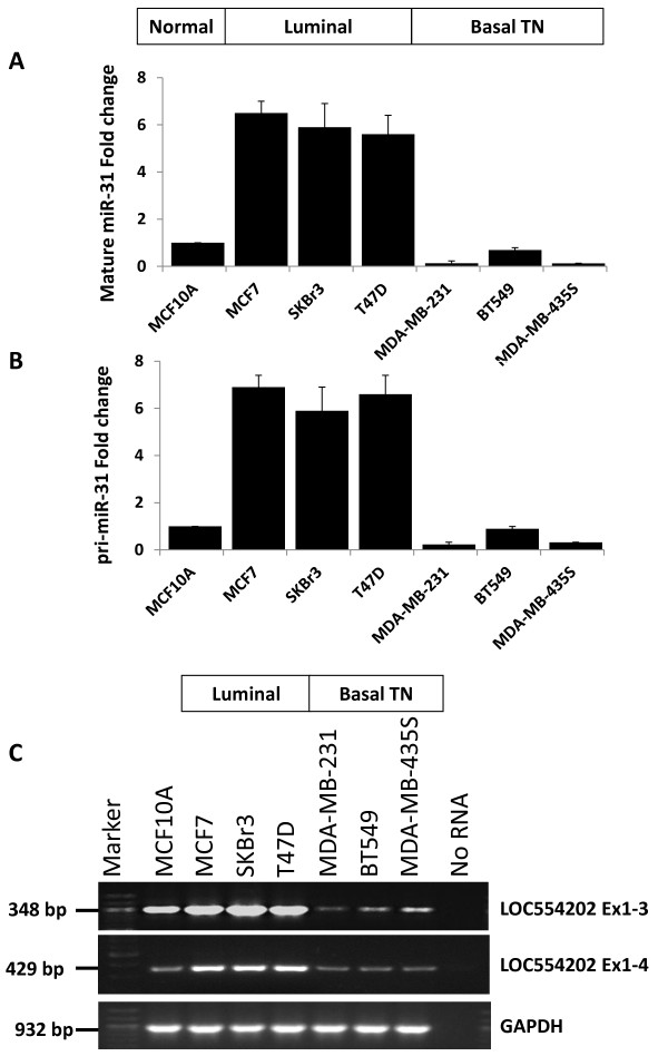
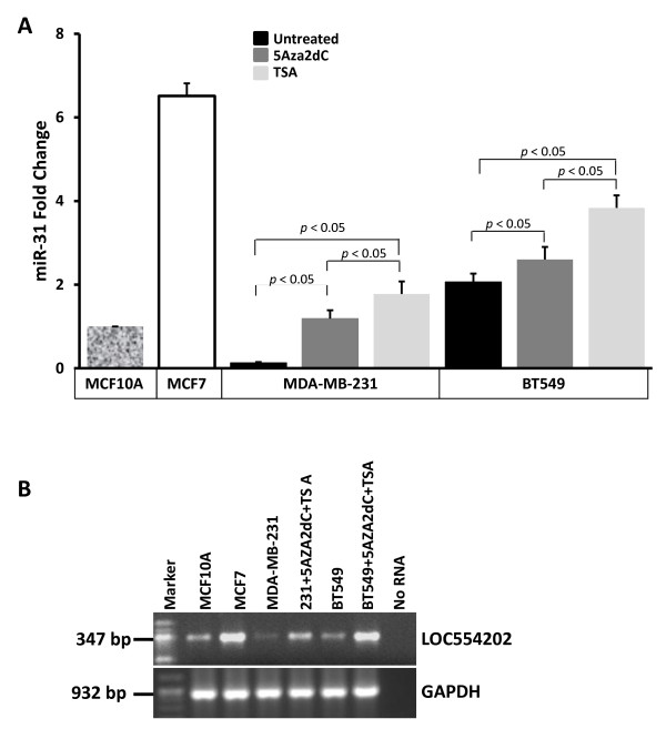
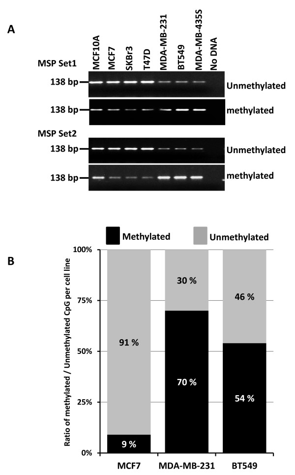
Results: In the present report, we identify promoter hypermethylation as one of the major mechanisms for silencing miR-31 in breast cancer, and in the triple-negative breast cancer (TNBC) cell lines of basal subtype, in particular. miR-31 maps to the intronic sequence of a novel long non-coding (lnc)RNA, LOC554202 and the regulation of its transcriptional activity is under control of LOC554202. Both miR-31 and the host gene LOC554202 are down-regulated in the TNBC cell lines of basal subtype and over-expressed in the luminal counterparts. Treatment of the TNBC cell lines with either a de-methylating agent alone or in combination with a de-acetylating agent resulted in a significant increase of both miR-31 and its host gene, suggesting an epigenetic mechanism for the silencing of these two genes by promoter hypermethylation. Finally, both methylation-specific PCR and sequencing of bisulfite-converted DNA demonstrated that the LOC554202 promoter-associated CpG island is heavily methylated in the TNBC cell lines and hypomethylated in the luminal subtypes.

Conclusion: Loss of miR-31 expression in TNBC cell lines is attributed to hypermethylation of its promoter-associated CpG island. Together, our results provide the initial evidence for a mechanism by which miR-31, an important determinant of the invasion metastasis cascade, is regulated in breast cancer.


Figures

Figure 1
Figure 1
Genomic organization of LCO554202/miR-31 locus and their associated CpG island. A) Schematic representation of the genomic organization of the LOC554202 gene. BAC clones RP11-344A7 and RP11-354P17 are shown with horizontal open bars. A strong CpG island spans exon 1 of LOC554202. miR-31 which maps within intron 1 of LOC554202 is shown with a red horizontal bar. The size of each exon and intron is also shown. The arrows below Exon 1, Exon 3 and Exon 4 represent the location and orientation of the primers used for RT-PCR of LOC554202. B) The location of the predicted CpG island (blue line) and LOC554202 exon 1 (green line) are shown with respect to the sequence of Human BAC clone RP11-344A7 on chromosome 9p21.2-22.3, Accession number AL137022, which contains the 5' end of the LOC554202, the host Gene of microRNA miR-31. Each vertical bar represents one CpG dinucleotide. Exon 1 of LOC554201 is shown as a red line. C) Schematic representation of the predicted CpG island and the location of the MSP primers sets and the bisulfite sequencing primers.
Figure 2
Figure 2
The genomic structure of the LOC554202 miR-31 is preserved in the BC cell lines used in this study. (A) Schematic representation of the mapping of the LOC554202 exons, the CpG island and miR-31 with respect to BAC clones RP11-344A7 and RP11-354P17. The mapping was confirmed by genomic PCR analysis. The size of the PCR product of each amplicon is shown on the left. ERVK6 was used as a positive control for the total genomic DNA. (B) Genomic PCR analysis of each exon of LOC554202 and miR-31 in the BC cell lines used in this study. The size of the PCR product of each amplicon is shown on the left. ERVK6 was used a positive control for the total genomic DNA.
Figure 3
Figure 3
miR-31 and its host gene LOC554202 are downregulated in basal TNBC cell lines and highly expressed in luminal non-invasive BC cell lines. (A and B) Quantitative real-time RT-PCR of the mature miR-31 (A) and the pri-miR-31 (B) in non-malignant breast epithelial MCF10A, luminal MCF7, SKBr3 and T47D, and basal triple-negative MDA-MB-435S, MDA-MB-231 and BT549 BC cell lines. miR-16 and RNUB6 were used for normalization. (C) Semi-quantitative RT-PCR of the LOC554202 transcript in the same cell lines. GAPDH was used an internal control.
Figure 4
Figure 4
miR-31 and LOC554202 are epigenetically regulated in breast cancer. (A) Quantitative real-time RT-PCR of miR-31 in the indicated cell lines before and after treatment with 5 Aza-2dC and TSA. miR-16 was used for normalization. (B) Semi-quantitative RT-PCR of LOC554202 after treatment of MDA-MB-231 and BT549 with the de-methylating 5Aza2dC and the de-acetylating Trichostatin A (TSA) agents. Expression of LOC554202 in the untreated cell lines is also shown. GAPDH was used as an internal control.
Figure 5
Figure 5
miR-31 promoter is heavily methylated in the basal TN versus the luminal BC cell lines. (A) Methylation specific PCR [MSP] of bisulfite-modified DNA of the indicated cell lines using 2 sets of primers. (B) Ratio of overall methylated over unmethylated CpGs by sequencing of bisulfite-modified DNA from the indicated cell lines. The location of the MSP and bisulfite sequencing primers is shown in Figure 1C.

References

    1. Berx G, Raspe E, Christofori G, Thiery JP, Sleeman JP. Pre-EMTing metastasis? Recapitulation of morphogenetic processes in cancer. Clin Exp Metastasis. 2007;24:587–597. doi: 10.1007/s10585-007-9114-6. - DOI - PubMed
    1. Chiang AC, Massague J. Molecular basis of metastasis. N Engl J Med. 2008;359:2814–2823. doi: 10.1056/NEJMra0805239. - DOI - PMC - PubMed
    1. Spaderna S, Schmalhofer O, Hlubek F, Jung A, Kirchner T, Brabletz T. Epithelial-mesenchymal and mesenchymal-epithelial transitions during cancer progression. Verh Dtsch Ges Pathol. 2007;91:21–28. - PubMed
    1. Nguyen DX, Bos PD, Massague J. Metastasis: from dissemination to organ-specific colonization. Nat Rev Cancer. 2009;9:274–284. doi: 10.1038/nrc2622. - DOI - PubMed
    1. May CD, Sphyris N, Evans KW, Werden SJ, Guo W, Mani SA. Epithelial-mesenchymal transition and cancer stem cells: a dangerously dynamic duo in breast cancer progression. Breast Cancer Res. 2011;13:202. - PMC - PubMed

Publication types

LinkOut - more resources