. 2012 Apr;99(4):202-8.
doi: 10.1016/j.ygeno.2012.01.005. Epub 2012 Jan 25.

Whole-exome sequencing in a single proband reveals a mutation in the CHST8 gene in autosomal recessive peeling skin syndrome

Affiliations

Whole-exome sequencing in a single proband reveals a mutation in the CHST8 gene in autosomal recessive peeling skin syndrome

Rita M Cabral et al. Genomics. 2012 Apr.

Abstract

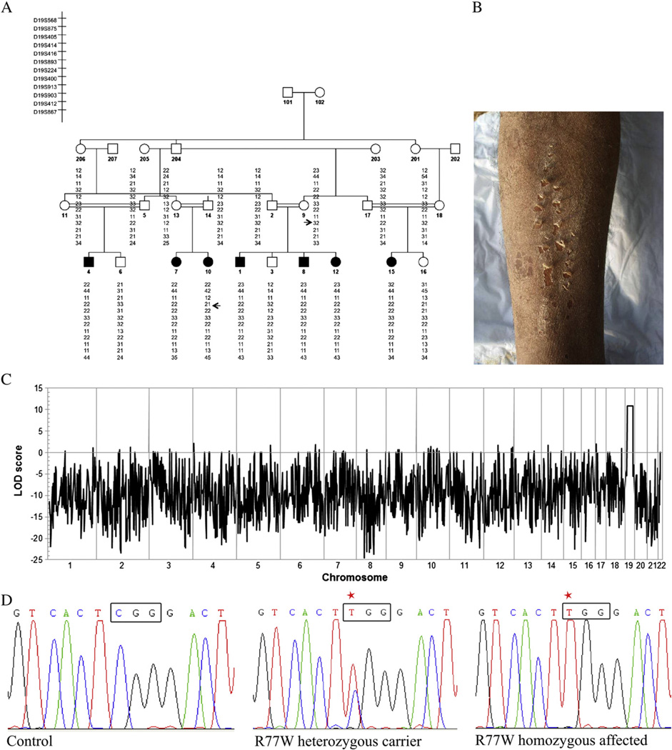
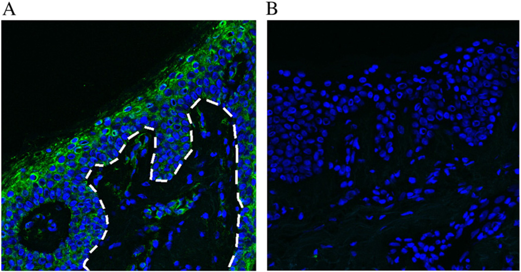
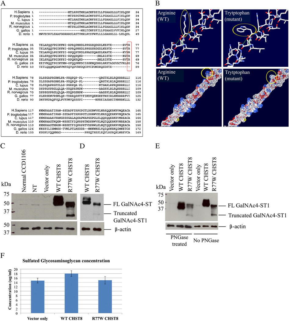
Generalized peeling skin syndrome (PSS) is an autosomal recessive genodermatosis characterized by lifelong, continuous shedding of the upper epidermis. Using whole-genome homozygozity mapping and whole-exome sequencing, we identified a novel homozygous missense mutation (c.229C>T, R77W) within the CHST8 gene, in a large consanguineous family with non-inflammatory PSS type A. CHST8 encodes a Golgi transmembrane N-acetylgalactosamine-4-O-sulfotransferase (GalNAc4-ST1), which we show by immunofluorescence staining to be expressed throughout normal epidermis. A colorimetric assay for total sulfated glycosaminoglycan (GAG) quantification, comparing human keratinocytes (CCD1106 KERTr) expressing wild type and mutant recombinant GalNAc4-ST1, revealed decreased levels of total sulfated GAGs in cells expressing mutant GalNAc4-ST1, suggesting loss of function. Western blotting revealed lower expression levels of mutant recombinant GalNAc4-ST1 compared to wild type, suggesting that accelerated degradation may result in loss of function, leading to PSS type A. This is the first report describing a mutation as the cause of PSS type A.


Figures

Fig. 1
Fig. 1
Autozygozity mapping and whole-exome sequencing identify homozygous missense mutation in CHST8 in a Pakistani family with PSS non-inflammatory type A. (A) Haplotype analysis in a large consanguineous family with PSS type A. Arrows indicate the key recombination events. (B) Clinical phenotype of an affected individual reveals superficial peeling of the skin. (C) Autozygozity mapping using the Affymetrix 10 K genotyping chip. A maximum LOD score of 10.9 was obtained for a 23 Mb region on chromosome 19. (D) Sanger sequencing of genomic DNA from patients 1–18 confirms presence of a homozygous c.229C>T mutation in affected individuals. Representative electropherograms for control, heterozygous carrier and homozygous affected individuals are shown (red star shows mutated nucleotide).
Fig. 2
Fig. 2
GalNAc4-ST1 is expressed throughout normal human epidermis. (A) Immunofluorescence staining of normal human frozen skin sections with a polyclonal antibody raised against GalNAc4-ST1 reveals expression of this protein throughout the epidermis, most predominantly in the granular and cornified layers. The dotted white line depicts the boundary between the epidermis and the dermis. (B) Negative control.
Fig. 3
Fig. 3
The c.229C>T (R77W) missense mutation results in decreased expression levels of full-length protein, loss of protein glycosylation and decreased activity of GalNAc4-ST1. (A) GalNAc4-ST1 protein sequence alignment across species (using HomoloGene, NCBI) shows that R77 is conserved among human, chimpanzee, dog, mouse and rat mammal species, but not in chicken or zebra fish. (B) Amino acid structure and electrostatic potential of GalNAc4-ST1 shows that the mutation perturbs amino acid hydrophilicity. Top panel: arginine (wild type) and tryptophan (mutant) amino acids are indicated with a red arrow. Blue = N, red = O, white = C, Bottom panel: a change in hydrophilicity due to the substitution of the basic amino acid arginine (white) by the non-polar amino acid tryptophan (blue) is indicated by the red arrow. Blue = basic amino acid, gray = non-polar amino acid. Figure generated using the Swiss-PdbViewer 4.0 application of the ExPASy Bioinformatics Resource Portal. (C and D) Western blot of whole cell lysates from CCD1106 keratinocytes transfected with c.229C>T mutant CHST8-pCMVscript (R77W CHST8) shows reduced levels of full length GalNAc4-ST1 (~49 kDa) compared to cells transfected with wild type CHST8-pCMVscript (WT CHST8). In addition, a lower molecular weight band (~35 kDa) is observed in whole lysates from cells overexpressing mutant CHST8, which is not detected in cells expressing wild type CHST8 and suggests increased degradation of mutant proteins. Normal keratinocytes (normal CCD1106), non-transfected keratinocytes that were incubated with Fugene HD only (NT) and cells transfected with empty pCMVscript (vector only) show low levels of full length GalNAc4-ST1. β-actin was used as a control of equal loading. FL = full length. (D) Lower exposure of blot B clearly showing lower levels of full length GalNAc4-ST1 in cells expressing mutant construct compared to wild type. (E) Western blot of wild type and mutant recombinant GalNAc4-ST1 proteins before and after treatment with PNGase reveals that, in contrast with wild type proteins, the mutant proteins are not subject to glycosylation. After PNGase treatment, WT proteins have smaller molecular weight than the untreated ones, but mutant proteins have identical molecular weights before and after treatment. (F) Colorimetric assay for total sulfated GAG quantification and comparison between cells transfected with wild type and mutant CHST8 constructs. Decreased levels of total sulfated GAGs are observed in cells expressing mutant GalNAc4-ST1 compared to wild type (n = 6), suggesting loss of function of mutant GalNAc4-ST1 proteins.

References

    1. Janjua SA, Hussain I, Khachemoune A. Facial peeling skin syndrome: a case report and a brief review. Int. J. Dermatol. 2007;46:287–289. - PubMed
    1. Köse O, Safali M, Koç E, Arca E, Açikgöz G, Ozmen I, Yeniay Y. Peeling skin diseases: 21 cases from Turkey and a review of the literature. J. Eur. Acad. Dermatol. Venereol. 2011 [Ahead of print]. - PubMed
    1. Bowden PE. Peeling skin syndrome: genetic defects in late terminal differentiation of the epidermis. J. Investig. Dermatol. 2011;3:561–564. - PubMed
    1. Ishida-Yamamoto A, Igawa S, Kishibe M. Order and disorder in corneocyte adhesion. J. Dermatol. 2011;7:645–654. - PubMed
    1. Telem DF, Israeli S, Sarig O, Sprecher E. Inflammatory peeling skin syndrome caused a novel mutation in CDSN. Arch. Dermatol. Res. 2011 [Ahead of print]. - PubMed

Publication types

MeSH terms

Supplementary concepts