Review
. 2012 Jun;121(3):235-50.
doi: 10.1007/s00412-012-0362-0.

Reconstituting the kinetochore–microtubule interface: what, why, and how

Affiliations
Review

Reconstituting the kinetochore–microtubule interface: what, why, and how

Bungo Akiyoshi et al. Chromosoma. 2012 Jun.

Abstract

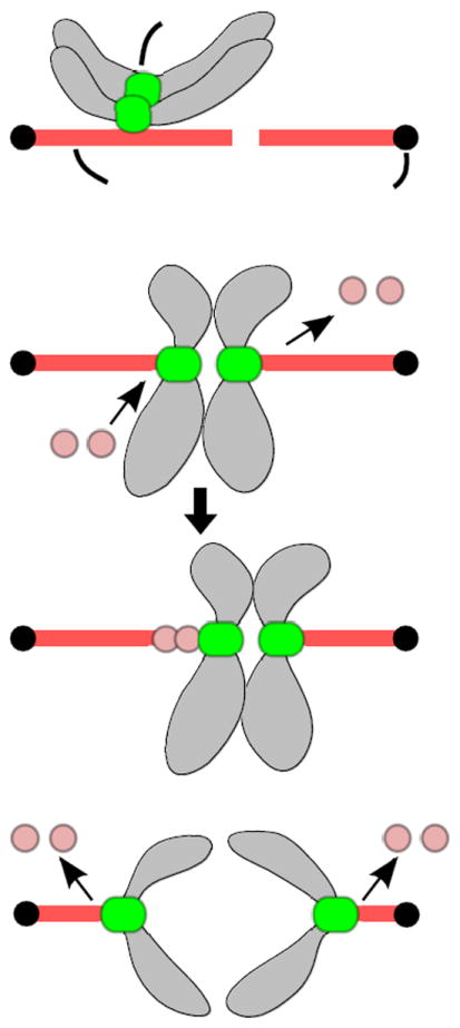
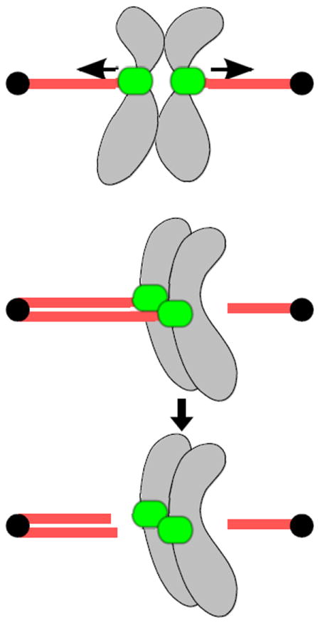
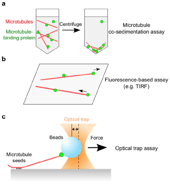
The kinetochore is the proteinaceous complex that governs the movement of duplicated chromosomes by interacting with spindle microtubules during mitosis and meiosis. Faithful chromosome segregation requires that kinetochores form robust load-bearing attachments to the tips of dynamic spindle microtubules, correct microtubule attachment errors, and delay the onset of anaphase until all chromosomes have made proper attachments. To understand how this macromolecular machine operates to segregate duplicated chromosomes with exquisite accuracy, it is critical to reconstitute and study kinetochore–microtubule interactions in vitro using defined components. Here, we review the current status of reconstitution as well as recent progress in understanding the microtubule-binding functions of kinetochores in vivo.


Figures

Fig. 1
Fig. 1
Lateral and end-on attachments. a Lateral attachments. Kinetochores initially encounter the lateral side of spindle microtubules during prometaphase. b End-on attachments during metaphase. Kinetochores interact with the tips of spindle microtubules where incorporation and dissociation of tubulin subunits occur. In this example, microtubules emanating from the left pole grow and those from the right pole shorten (top), leading to rightward movement of the attached chromosomes (bottom). c End-on attachments during anaphase. Linkage between sister chromatids is lost and microtubules from both sides shorten, resulting in the separation of sister chromatids to opposite poles.
Fig. 2
Fig. 2
Different types of kinetochore-microtubule attachments. a Bi-oriented attachments in which sister kinetochores attach to microtubules emanating from opposite poles. Kinetochores are under robust tension due to microtubule pulling forces that are opposed by linkage between sister chromatids. Tension stabilizes these proper bi-oriented attachments. b Mono-oriented attachment in which sister kinetochores are attached to microtubules emanating from the same pole (top). Lack of tension destabilizes these improper attachments, giving cells another chance to achieve correct bi-oriented attachments (bottom).
Fig. 3
Fig. 3
Examples of microtubule-binding assays. a A microtubule co-sedimentation assay analyzes whether proteins of interest stay associated with stabilized (non-dynamic) microtubules that are pelleted by centrifugation. The level of co-sedimentation is typically determined by immunoblots. b A fluorescence-based assay observes binding between fluorescent microtubules attached to a coverslip and fluorescently labeled proteins. TIRF microscopy enables visualization of molecules only in the evanescent field (within ~100 nm of cover slip), providing single-molecule sensitivity (i.e. observation of binding events between individual microtubules and individual kinetochore proteins or complexes). Dynamically growing and shortening microtubules can also be used, allowing studies of lateral versus end-on attachments. c Optical trap assays use a focused laser beam to apply force to the interaction between bead-bound proteins and dynamic microtubule tips. Growth and shrinkage of individual microtubules can be monitored by video-enhanced microscopy and the number of bead-bound proteins can be diluted low enough to allow studies of single molecules or complexes.
Fig. 4
Fig. 4
Effects of tension on microtubule tip dynamics and attachment stability. a End-on attachments allow kinetochores and tension to directly regulate microtubule tip dynamics. b Tension stabilizes reconstituted kinetochore-microtubule interactions. Left: Tension increases rescue rate and decreases catastrophe rates, resulting in a state where microtubules spend more time in the assembly phase. Right: There is also a tendency for the detachment rate from shortening microtubules to decrease in the presence of higher tension whereas that from growing microtubules increases. Changes in these four parameters result in a net increase in total attachment time as tension is increased within a certain force range (0.5 – 5 pN) (Akiyoshi et al. 2010).

References

    1. Akiyoshi B, Nelson CR, Ranish JA, Biggins S. Quantitative proteomic analysis of purified yeast kinetochores identifies a PP1 regulatory subunit. Genes Dev. 2009a;23:2887–2899. doi: 10.1101/gad.1865909. - DOI - PMC - PubMed
    1. Akiyoshi B, Nelson CR, Ranish JA, Biggins S. Analysis of Ipl1-mediated phosphorylation of the Ndc80 kinetochore protein in Saccharomyces cerevisiae. Genetics. 2009b;183:1591–1595. doi: 10.1534/genetics.109.109041. - DOI - PMC - PubMed
    1. Akiyoshi B, Sarangapani KK, Powers AF, et al. Tension directly stabilizes reconstituted kinetochore-microtubule attachments. Nature. 2010;468:576–579. doi: 10.1038/nature09594. - DOI - PMC - PubMed
    1. Al-Bassam J, Chang F. Regulation of microtubule dynamics by TOG-domain proteins XMAP215/Dis1 and CLASP. Trends Cell Biol. 2011;21:604–614. doi: 10.1016/j.tcb.2011.06.007. - DOI - PMC - PubMed
    1. Allshire RC, Karpen GH. Epigenetic regulation of centromeric chromatin: old dogs, new tricks? Nat Rev Genet. 2008;9:923–937. doi: 10.1038/nrg2466. - DOI - PMC - PubMed

Publication types

MeSH terms

LinkOut - more resources