

Cerebral and muscle MRI abnormalities in myotonic dystrophy
- PMID: 22290140
- PMCID: PMC3350604
- DOI: 10.1016/j.nmd.2012.01.003
Cerebral and muscle MRI abnormalities in myotonic dystrophy
Abstract
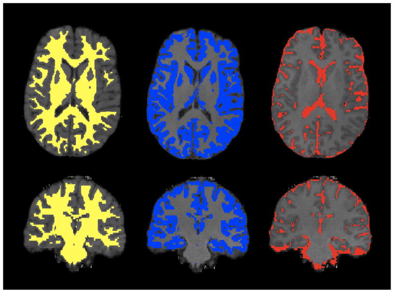
Pathophysiological mechanisms underlying the clinically devastating CNS features of myotonic dystrophy (DM) remain more enigmatic and controversial than do the muscle abnormalities of this common form of muscular dystrophy. To better define CNS and cranial muscle changes in DM, we used quantitative volumetric and diffusion tensor MRI methods to measure cerebral and masticatory muscle differences between controls (n=5) and adults with either congenital (n=5) or adult onset (n=5) myotonic dystrophy type 1 and myotonic dystrophy type 2 (n=5). Muscle volumes were diminished in DM1 and strongly correlated with reduced white matter integrity and gray matter volume. Moreover, correlation of reduced fractional anisotropy (white matter integrity) and gray matter volume in both DM1 and DM2 suggests that these abnormalities may share a common underlying pathophysiological mechanism. Further quantitative temporal and spatial characterization of these features will help delineate developmental and progressive neurological components of DM, and help determine the causative molecular and cellular mechanisms.
Copyright © 2012 Elsevier B.V. All rights reserved.
Figures


References
-
- Brook JD, McCurrach ME, Harley HG, et al. Molecular basis of myotonic dystrophy: expansion of a trinucleotide (CTG) repeat at the 3′ end of a transcript encoding a protein kinase family member. Cell. 1992;68:799–808. - PubMed
-
- Fu YH, Pizzuti A, Fenwick RG, Jr, et al. An unstable triplet repeat in a gene related to myotonic muscular dystrophy. Science. 1992;255:1256–1258. - PubMed
-
- Mahadevan M, Tsilfidis C, Sabourin L, et al. Myotonic dystrophy mutation: an unstable CTG repeat in the 3′ untranslated region of the gene. Science. 1992;255:1253–1255. - PubMed
-
- Liquori CL, Ricker K, Moseley ML, et al. Myotonic dystrophy type 2 caused by a CCTG expansion in intron 1 of ZNF9. Science. 2001;293:864–867. - PubMed
-
- Ranum LP, Rasmussen PF, Benzow KA, Koob MD, Day JW. Genetic mapping of a second myotonic dystrophy locus. Nat Genet. 1998;19:196–198. - PubMed
Publication types
MeSH terms
Grants and funding
LinkOut - more resources
Full Text Sources
