Review
. 2012 Jun;5(3):264-73.
doi: 10.1007/s12265-012-9349-8.

Elastin in large artery stiffness and hypertension

Affiliations
Review

Elastin in large artery stiffness and hypertension

Jessica E Wagenseil et al. J Cardiovasc Transl Res. 2012 Jun.

Abstract

Large artery stiffness, as measured by pulse wave velocity, is correlated with high blood pressure and may be a causative factor in essential hypertension. The extracellular matrix components, specifically the mix of elastin and collagen in the vessel wall, determine the passive mechanical properties of the large arteries. Elastin is organized into elastic fibers in the wall during arterial development in a complex process that requires spatial and temporal coordination of numerous proteins. The elastic fibers last the lifetime of the organism but are subject to proteolytic degradation and chemical alterations that change their mechanical properties. This review discusses how alterations in the amount, assembly, organization, or chemical properties of the elastic fibers affect arterial stiffness and blood pressure. Strategies for encouraging or reversing alterations to the elastic fibers are addressed. Methods for determining the efficacy of these strategies, by measuring elastin amounts and arterial stiffness, are summarized. Therapies that have a direct effect on arterial stiffness through alterations to the elastic fibers in the wall may be an effective treatment for essential hypertension.


Figures

Figure 1
Figure 1
Representative circumferential stress-stretch relationship for the mouse ascending aorta (data from Carta et al. [87]). Other large elastic arteries from different vertebrate animals show similar behavior. The low pressure, low stretch region dominated by elastin and the high pressure, high stretch region dominated by collagen are shown. At normal blood pressures, the regions overlap in the physiologic range.
Figure 2
Figure 2
Elastic fiber assembly in E14 mouse aorta. Electron micrograph of the inner elastic lamina between the endothelial cells (EC) at the luminal surface of the vessel and the first layer of SMCs in the vessel wall. Elastin aggregates (arrow) associate with microfibrils (arrowhead) near the cell surface.
Figure 3
Figure 3
Left ventricular blood pressure and aortic stiffness in WT and Eln-/- mice (data complied from Wagenseil et al. [42,41]). There are no differences in systolic pressure at E18, but by P1 the pressure in Eln-/- mice is almost double that of WT (A). The incremental stiffness of the ascending aorta is significantly increased in Eln-/- mice for at least two pressure steps at E18 (B) and P1 (C). In both cases, the stiffness differences are in the low to mid pressure range in the region dominated by elastin. The aortic stiffness increases with pressure as more collagen fibers are recruited. * = p <.05 between WT and Eln-/- by unpaired t-test with unequal distributions.
Figure 4
Figure 4
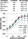
Blood pressure and arterial stiffness are inversely related to elastin amounts (data compiled from Faury et al. [88], Hirano et al. [50] and Wagenseil et al. [46]). Approximate elastin percentage and systolic pressure (SP) are shown for each mouse genotype (top). The pressure increases as the elastin amounts decrease. Diameter-pressure curves for the left common carotid artery (bottom). The curves become more horizontal, increasing in stiffness, as elastin amounts decrease. The 30 and 60% systolic pressure and diameter-pressure curves are significantly different (p < .05) from 100%.
Figure 5
Figure 5
Possible targets for therapeutic strategies to alter elastic fibers and affect arterial stiffness and blood pressure. Stiffness of the elastic fibers is modified through crosslinking and calcification. Assembly of the elastic fibers depends on synthesis of elastin and associated proteins. Degradation of the elastic fibers is controlled through protease and anti-protease activity.
Figure 6
Figure 6
Pressure-diameter, force-length diagram for a cylindrical artery specimen. The artery is mounted on hollow cannulae at the unloaded length (L) and diameter (D). The artery is stretched to some length (z) and inflated with fluid pressure through the cannulae to some diameter (d). Reproduced with permission from Christopher Pozzo.

References

    1. Mulvany MJ. Small artery remodelling in hypertension: causes, consequences and therapeutic implications. Med Biol Eng Comput. 2008;46(5):461–467. doi:10.1007/s11517-008-0305-3. - PubMed
    1. Kearney PM, Whelton M, Reynolds K, Muntner P, Whelton PK, He J. Global burden of hypertension: analysis of worldwide data. Lancet. 2005;365(9455):217–223. doi:10.1016/S0140-6736(05)17741-1. - PubMed
    1. Cecelja M, Chowienczyk P. Dissociation of aortic pulse wave velocity with risk factors for cardiovascular disease other than hypertension: a systematic review. Hypertension. 2009;54(6):1328–1336. doi:HYPERTENSIONAHA.109.137653 [pii] 10.1161/HYPERTENSIONAHA.109.137653. - PubMed
    1. Messerli FH, Frohlich ED, Ventura HO. Arterial compliance in essential hypertension. J Cardiovasc Pharmacol. 1985;7(Suppl 2):S33–35. - PubMed
    1. Faury G, Maher GM, Li DY, Keating MT, Mecham RP, Boyle WA. Relation between outer and luminal diameter in cannulated arteries. Am J Physiol. 1999;277(5 Pt 2):H1745–1753. - PubMed

Publication types

MeSH terms