Review
. 2012 Jan 31;9(6):319-32.
doi: 10.1038/nrcardio.2012.3.

Potassium-channel mutations and cardiac arrhythmias--diagnosis and therapy

Affiliations
Review

Potassium-channel mutations and cardiac arrhythmias--diagnosis and therapy

John R Giudicessi et al. Nat Rev Cardiol. .

Abstract

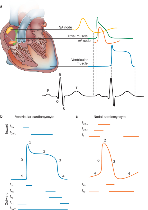
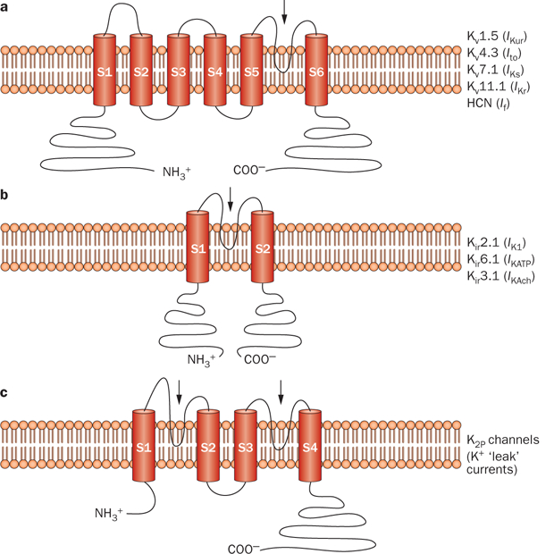
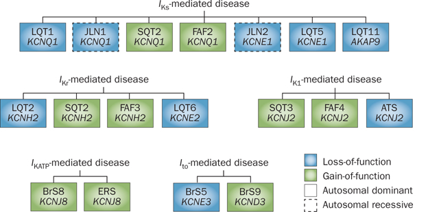
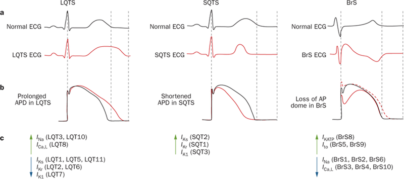
The coordinated generation and propagation of action potentials within cardiomyocytes creates the intrinsic electrical stimuli that are responsible for maintaining the electromechanical pump function of the human heart. The synchronous opening and closing of cardiac Na(+), Ca(2+), and K(+) channels corresponds with the activation and inactivation of inward depolarizing (Na(+) and Ca(2+)) and outward repolarizing (K(+)) currents that underlie the various phases of the cardiac action potential (resting, depolarization, plateau, and repolarization). Inherited mutations in pore-forming α subunits and accessory β subunits of cardiac K(+) channels can perturb the atrial and ventricular action potential and cause various cardiac arrhythmia syndromes, including long QT syndrome, short QT syndrome, Brugada syndrome, and familial atrial fibrillation. In this Review, we summarize the current understanding of the molecular and cellular mechanisms that underlie K(+)-channel-mediated arrhythmia syndromes. We also describe translational advances that have led to the emerging role of genetic testing and genotype-specific therapy in the diagnosis and clinical management of individuals who harbor pathogenic mutations in genes that encode α or β subunits of cardiac K(+) channels.


Conflict of interest statement

Competing interests

M. J. Ackerman declares associations with the following companies: Biotronik, Boston Scientific, Medtronic, St. Jude Medical, and Transgenomic. See the article online for full details of the relationships. J. R. Giudicessi declares no competing interests.

Figures

Figure 1 |
Figure 1 |
Normal electrical activity of the heart. a | Schematic representation of the cardiac conduction system and the correlation between the action potentials of cardiomyocytes in distinct regions of the heart and the surface electrocardiogram. b | Temporal relationship between the inward and outward currents that contribute to the distinct phases (0–4) of the ventricular cardiomyocyte action potential. c | Temporal relationship between the inward and outward currents that contribute to the distinct phases (0–4) of the nodal cardiomyocyte action potential (which lacks a defined phase 1). Abbreviations: AV, atrioventricular; ICa,L, inward depolarizing Ca2+ current (through L-type Ca2+ channels); ICa,T, inward depolarizing Ca2+ current (through T-type Ca2+ channels); If, inward-rectifier mixed Na+ and K+ ‘funny’ current; IK1, inward-rectifier K+ current; IKATP, ATP-sensitive K+ current; IKr, rapid component of the delayed-rectifier K+ current; IKs, slow component of the delayed-rectifier K+ current; INa, inward depolarizing Na+ current; Ito, transient outward K+ current; SA, sinoatrial.
Figure 2 |
Figure 2 |
Predicted protein topology of cardiac K+-channel α subunits. Schematic representations of a | the six-transmembrane one-pore-region voltage-dependent K+-channel (Kv) α subunits that conduct IKur, Ito, IKs, IKr, and If in the heart, b | the two-transmembrane one-pore-region inward-rectifying K+-channel (Kir) α subunits that conduct IK1, IKATP, and IKAch in the heart, and c | the four-transmembrane two-pore-region K+ channel (K2P) responsible for ‘leak’ K+ currents. The arrows indicate the location of the pore-forming region(s). Abbreviations: HCN, hyperpolarization-activated cyclic-nucleotide-gated channel; If, inward-rectifier mixed Na+ and K+ ‘funny’ current; IK1, inward-rectifier K+ current; IKAch, acetylcholine-activated inward-rectifier K+ current; IKATP, ATP-sensitive K+ current; IKr, rapid component of the delayed-rectifier K+ current; IKs, slow component of the delayed-rectifier K+ current; IKur, ultra-rapid component of the delayed-rectifier K+ current; Ito, transient outward K+ current.
Figure 3 |
Figure 3 |
Classification of cardiac K+ channelopathies stemming from mutations in α or β subunits on the basis of currents and channels. Current-centric depiction of the K+ channelopathies arising from loss-of-function (blue boxes) or gain-of-function (green boxes) mutations that perturb IKs, IKr, IK1, IKATP, or Ito. Abbreviations: BrS, Brugada syndrome; ERS, early repolarization syndrome; FAF, familial atrial fibrillation; I, inward-rectifier K+ current; I, ATP-sensitive K+ current; I, rapid component of the delayed-rectifier K+ current; IKs, slow component of the delayed-rectifier K+ current; Ito, transient outward K+ current; JLN, Jervell and Lange–Nielsen syndrome; LQT, long QT syndrome; SQT, short QT syndrome. Permission obtained from Wolters Kluwer Health © Priori et al. Inherited arrhythmogenic disease: the complexity beyond monogenic disorders. Circ. Res. 94(2), 140–145 (2004).
Figure 4 |
Figure 4 |
Pathophysiological mechanisms of K+-channel-mediated arrhythmias at the ionic and cellular level. a | Schematic representation of a normal ECG (black) and typical ECGs for patients with LQTS (left, red), SQTS (middle, red), and BrS (right, red), illustrating QT lengthening, QT shortening, and coved ST-segment elevation in the right precordial leads V1–V3 and presence of a J-wave, respectively. b | Tracings of normal ventricular action potentials (black) and tracings that display epicardial action-potential prolongation in LQTS (left, red), action-potential abbreviation in SQTS (middle, red), and the transmural gradient between the epicardial action potential (right, solid red line) and endocardial action-potential (right, dotted red line) that results in the inscription of the J-wave in BrS. c | Perturbations in ion currents that contribute to the pathogenesis of LQTS, SQTS, and BrS. Green upwards arrows indicate gain-of-function mutations, whereas blue downwards arrows represent loss-of-function mutations. Abbreviations: AP, action potential; APD, action-potential duration; BrS, Brugada syndrome; ECG, electrocardiogram; LQT or LQTS, long QT syndrome; SQT or SQTS, short QT syndrome. Reproduced from Wilde, A. A. & Bezzina, C. R. Genetics of cardiac arrhythmias. Heart 91(10), 1352–1358 © 2005, with permission from BMJ Publishing Group Ltd.
Figure 5 |
Figure 5 |
The spectrum of QTc values and associated clinical indications for adults with a QTc value in the prolonged, borderline-prolonged, normal, borderline-short, and extremely short ranges. This Figure represents a modification of the algorithm initially proposed by G. M. Vincent and later modified by S. Viskin. Abbreviations: ECG, electrocardiogram; LQTS, long QT syndrome; QTc, heart-rate-corrected QT interval; SQTS, short QT syndrome. Reprinted from Viskin, S. The QT interval: too long, too short or just right. Heart Rhythm 6(5), 711–715 © 2009, with permission from the Heart Rhythm Society.
Figure 6 |
Figure 6 |
The probabilistic nature of LQTS genetic testing. Depicted is the probability of pathogenicity for mutations that localize to the N-terminal, transmembrane, or C-terminal regions of the Kv7.1 and Kv11.1 channels that collectively constitute the two major causes of congenital LQTS. Although radical mutations have a >90% probability of being pathogenic, the probability for missense mutations varies depending on the location within each respective K+ channel. Missense mutations in areas shaded in red carry a high probability (>80%), mutations in yellow have an intermediate probability (51–80%), and the pathogenicity of mutations in blue areas is uncertain (<50%). Abbreviations: cNDB, cyclic-nucleotide-binding domain; LQTS, long QT syndrome; PAC, PAS-associated C-terminal domain; PAS, Per–Arnt–Sim domain; SAD, subunit assembly domain. Permission obtained from Wolters Kluwer Health © Tester, D. J. & Ackerman, M. J. Genetic testing for potentially lethal, highly treatable inherited cardiomyopathies/channelopathies in clinical practice. Circulation 123(9), 1021–1037 (2011).

References

    1. Nerbonne JM & Kass RS Molecular physiology of cardiac repolarization. Physiol. Rev 85, 1205–1253 (2005). - PubMed
    1. Wang Q et al. Positional cloning of a novel potassium channel gene: KVLQT1 mutations cause cardiac arrhythmias. Nat. Genet 12, 17–23 (1996). - PubMed
    1. Curran ME et al. A molecular basis for cardiac arrhythmia: HERG mutations cause long QT syndrome. Cell 80, 795–803 (1995). - PubMed
    1. Plaster NM et al. Mutations in Kir2.1 cause the developmental and episodic electrical phenotypes of Andersen’s syndrome. Cell 105, 511–519 (2001). - PubMed
    1. Hedley PL et al. The genetic basis of Brugada syndrome: a mutation update. Hum. Mutat 30, 1256–1266 (2009). - PubMed

Publication types

MeSH terms

Substances