Review
. 2012 Jan 31;8(4):212-27.
doi: 10.1038/nrendo.2011.234.

MicroRNA control of bone formation and homeostasis

Affiliations
Review

MicroRNA control of bone formation and homeostasis

Jane B Lian et al. Nat Rev Endocrinol. .

Abstract

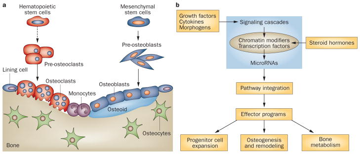
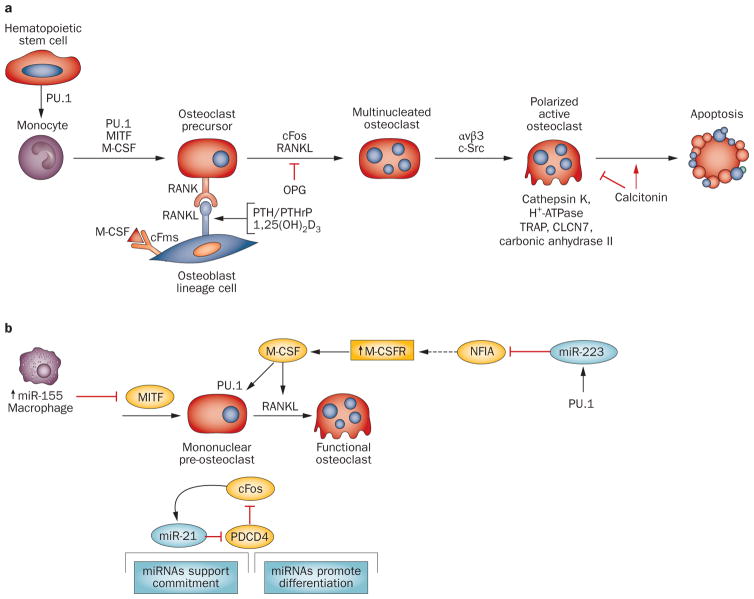
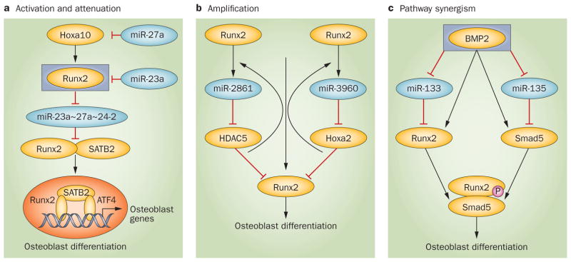
MicroRNAs (miRNAs) repress cellular protein levels to provide a sophisticated parameter of gene regulation that coordinates a broad spectrum of biological processes. Bone organogenesis is a complex process involving the differentiation and crosstalk of multiple cell types for formation and remodeling of the skeleton. Inhibition of mRNA translation by miRNAs has emerged as an important regulator of developmental osteogenic signaling pathways, osteoblast growth and differentiation, osteoclast-mediated bone resorption activity and bone homeostasis in the adult skeleton. miRNAs control multiple layers of gene regulation for bone development and postnatal functions, from the initial response of stem/progenitor cells to the structural and metabolic activity of the mature tissue. This Review brings into focus an emerging concept of bone-regulating miRNAs, the evidence for which has been gathered largely from in vivo mouse models and in vitro studies in human and mouse skeletal cell populations. Characterization of miRNAs that operate through tissue-specific transcription factors in osteoblast and osteoclast lineage cells, as well as intricate feedforward and reverse loops, has provided novel insights into the supervision of signaling pathways and regulatory networks controlling normal bone formation and turnover. The current knowledge of miRNAs characteristic of human pathologic disorders of the skeleton is presented with a future goal towards translational studies.


Conflict of interest statement

Competing interests

The authors declare no competing interests.

Figures

Figure 1
Figure 1
The bone remodeling cycle and regulation of bone tissue homeostasis. a | Cellular activities supporting bone remodeling. A remodeling cycle, regulated by parathyroid hormone and 1,25-dihydroxyvitamin D3, is initiated with a resorption phase by activated osteoclasts that solubilize bone mineral and degrade the matrix. Osteoclasts originate from hematopoietic stem cells which differentiate first (dotted arrow) to a committed mononuclear preosteoclast cell that fuses to form the multinucleated cells. Attachment of osteoclasts to the bone surface creates a local acidic environment forming a resorption pit. The activities of monocytes or macrophages remove debris (reversal phase), followed by a bone formation phase by osteoblasts, producing osteoid matrix which will mineralize. Growth factors are released from the bone matrix during resorption, which increases the preosteoblast population to replace eroded bone surfaces. b | Regulation of bone programs. The skeletal landscape is illustrated as an integration of genetic (expressed genes) and epigenetic (microRNAs [miRNA]) factors that leads to the effector programs to support the three major activities of bone (lower boxes). The integration of these regulators is facilitated by post-transcriptional control that appears to be highly regulated by miRNAs.
Figure 2
Figure 2
The osteoblast differentiation program. a | In vivo: bone surface shows organization of indicated osteoblast lineage cells (black, mineralized tissue). Mesenchymal stem cells and osteoprogenitor cells cannot be seen. b | In vitro: stages of differentiation of committed preosteoblast cells isolated from newborn rodent calvarium or bone marrow stromal cells. Peak expression of genes that are markers for the three major stages are shown. At mineralization, a feedback signal from sclerostin secreted by osteocytes inhibits BMP and Wnt osteogenic-mediated bone formation by regulating the number of cells entering the osteoblast lineage. c | Examples of transcription factors regulating osteoblast differentiation and in vivo bone formation are shown. Within the triangle are those that increase during differentiation, whereas those above the triangle are functional on gene promoters at the indicated stages of maturation. Permission obtained from American Society for Bone and Mineral Research © Favus, M. J. (Ed.) Primer on the Metabolic Bone Diseases and Disorders of Mineral Metabolism, 6th edn (2006).
Figure 3
Figure 3
Osteoclast differentiation sequence and effect of microRNAs. a | Stages in the differentiation of the multinucleated osteoclast from its hematopoietic precursor are illustrated with key transcription factors and regulatory proteins established as critical for progression to the activated osteoclast. These include the RANK–RANKL interaction regulated by the indicated hormones and the inhibitor of RANK signaling osteoprotegerin. Integrin (αvβ3) mediates attachment of osteoclasts to bone surface and c-Src signaling induces polarization of the osteoclast and formation of the characteristic ruffled border for active bone resorption. b | MicroRNAs regulating commitment to osteoclastogenesis have been identified. Indicated are three different mechanisms. miR-155 functions as an inhibitor of osteoclastogenesis, being highly expressed in macrophages to support robust expression of this phenotype by inhibiting MITF, essential for preosteoclast differentiation. PU.1 initiates a feedforward mechanism increasing miR-223 which downregulates an inhibitor of osteoclast differentiation, NFIA, resulting in an increase in M-CSFR and thereby M-CSF functional activity. Also shown is a regulatory loop between miR-21 and cFos (AP-1), which activates many osteoclast genes essential for multinucleated cell formation and promotes resorptive activity. Abbreviations: αvβ3, integrin αvβ3; CLNC7, chloride channel; OPG, osteoprotegerin; TRAP, tartrate-resistant acid phosphatase.
Figure 4
Figure 4
Effect of microRNAs (miRNAs) on osteoblast differentiation. a | BMP2 induces mesenchymal stromal cell commitment to osteogenesis. Below the subpopulations of osteoblasts (top row) are boxes indicating the major activities of miRNAs influencing the continuous maturation of the cells from the mesenchymal stromal cells to the osteocyte. Selected miRNAs (blue) in relation to their targets (yellow) influencing each stage to regulate progression of differentiation are indicated. Note that the miR-29 family targets several inhibitors of Wnt, MAPK signaling and collagens at different stages of differentiation., Other miRNAs, such as miR-335, also target Wnt inhibitors. b | The consequence of Dicer deletion in osteoblasts and osteocytes (left panel; Dicer-C/C) driven by osteocalcin (OC-Cre) (right panel; Dicer ΔOC/ΔOC). Loss-of-function of mature miRNAs in this population results in increased bone mass potentially by relieving repression of Runx2 miRNAs (n=11) and collagen protein levels. Courtesy of J. B. Lian, University of Massachusetts Medical School, USA.
Figure 5
Figure 5
Regulatory circuits operating in osteoblast lineage cells. a | Activation and attenuation pathways. From top, BMP2 induces Hoxa10 which activates Runx2., Runx2 downregulates expression of the cluster miR23a~27a~24-2 directly through a Runx regulatory element in the promoter for this cluster, which results in increased Runx2 and its coregulator SATB2 protein, that are targets of the cluster miRNAs. Runx2, SATB2 and ATF4 form an activating complex that is essential for differentiation. The cluster becomes robustly elevated at mineralization through a feedback mechanism by which miR-27a suppresses Hoxa10 and miR-23a attenuates Runx2 protein. This feedforward and feed reverse circuit exemplifies the central role of this microRNA cluster in promoting and attenuating the progression of differentiation to regulate bone formation. b | Amplification mechanism. Two microRNAs form the cluster miR-2861~miR-3960, each of which targets inhibitors of Runx2 activity, HDAC5 and Hoxa2, respectively. This downregulation increases Runx2 transcriptional activity. The effect is amplified because Runx2 transactivates the cluster through a Runx2 binding site in the cluster’s promoter. Thus, a regulatory circuit exists where Runx2 and a microRNA cluster operate in a coordinate manner. c | Pathway synergism. BMP2 downregulates miRNAs that suppress two essential factors for osteogenesis that form a transcriptional complex. The integration of Runx2 and miRNAs occurs at two levels—Runx2 regulation of miRNA expression and miRNA blockade of Runx2 inhibitors.
Figure 6
Figure 6
Allocation of mesenchymal stem cells (MSCs) to lineage-specific phenotypes by transcription factors and microRNAs. Schematic illustration of MSC lineages directed by cell-type specific transcription factors (arrows). Selected miRNAs highly expressed in MSCs are shown because they are downregulated during differentiation into phenotype-committed cells. Cell-type-related miRNAs targeting the transcription factors or their coregulatory proteins are indicated. Although the transcription factors are attenuated by miRNAs at different stages of maturation, they are critical for regulating a normal program of differentiation and for cell specification within a tissue. Relevant references to support this concept for the indicated tissues are as follows: muscle,,, nerve,–, fat,,,,, bone and cartilage (Table 1). *Many miRNAs repress Runx2 directly (shown in Figure 7), and to date three miRNAs have been shown to repress osterix: miR-125b in vascular smooth muscle cells prevents calcification and miR-138 in MSCs retains stemness, and miR-637 is expressed in adipocytes.
Figure 7
Figure 7
MicroRNAs (miRNAs) regulating Runx2. a | The mouse Runx2 3′ untranslated region. Binding sites are shown for validated miRNAs., MiRNAs are expressed at high (red), moderate (black) or low (blue) levels in murine MC3T3 preosteoblasts (at confluency in growth media). In addition, miR-335 targets the human Runx2 3′ untranslated region. b | Modification of Runx2 miRNAs during differentiation. The osteochondral progenitor is a bipotent cell and commitment to either the osteoblastic or chondrocytic lineage is regulated in part by miRNAs. A subset of Runx2-targeting miRNAs is downregulated as cells are induced into osteogenesis, while a subset is upregulated in differentiated osteocytes to support physiological regulation of Runx2. A group of miRNAs targeting Runx2 is highly expressed at the onset of chondrogenesis to inhibit the bone phenotype. However, with further differentiation to the hypertrophic chondrocyte, miRNAs targeting Runx2 are downregulated.

References

    1. Del Fattore A, Teti A, Rucci N. Osteoclast receptors and signaling. Arch Biochem Biophys. 2008;473:147–160. - PubMed
    1. Ross FP, Teitelbaum SL. αvβ3 and macrophage colony-stimulating factor: partners in osteoclast biology. Immunol Rev. 2005;208:88–105. - PubMed
    1. Miyazaki T, Tanaka S, Sanjay A, Baron R. The role of c-Src kinase in the regulation of osteoclast function. Mod Rheumatol. 2006;16:68–74. - PubMed
    1. Edwards CM, Mundy GR. Eph receptors and ephrin signaling pathways: a role in bone homeostasis. Int J Med Sci. 2008;5:263–272. - PMC - PubMed
    1. Sims NA, Gooi JH. Bone remodeling: Multiple cellular interactions required for coupling of bone formation and resorption. Semin Cell Dev Biol. 2008;19:444–451. - PubMed

Publication types