. 2012 Apr;32(7):1312-20.
doi: 10.1128/MCB.06222-11. Epub 2012 Jan 30.

The chromatin-remodeling enzymes BRG1 and CHD4 antagonistically regulate vascular Wnt signaling

Affiliations

The chromatin-remodeling enzymes BRG1 and CHD4 antagonistically regulate vascular Wnt signaling

Carol D Curtis et al. Mol Cell Biol. 2012 Apr.

Abstract

Canonical Wnt signaling plays an important role in embryonic and postnatal blood vessel development. We previously reported that the chromatin-remodeling enzyme BRG1 promotes vascular Wnt signaling. Vascular deletion of Brg1 results in aberrant yolk sac blood vessel morphology, which is rescued by pharmacological stimulation of Wnt signaling with lithium chloride (LiCl). We have now generated embryos lacking the chromatin-remodeling enzyme Chd4 in vascular endothelial cells. Unlike Brg1 mutants, Chd4 mutant embryos had normal yolk sac vascular morphology. However, concomitant deletion of Chd4 and Brg1 rescued vascular abnormalities seen in Brg1 mutant yolk sacs to the same extent as LiCl treatment. We hypothesized that Wnt signaling was upregulated in Chd4 mutant yolk sac vasculature. Indeed, we found that Chd4 deletion resulted in upregulation of the Wnt-responsive transcription factor Tcf7 and an increase in Wnt target gene expression in endothelial cells. Furthermore, we identified one Wnt target gene, Pitx2, that was downregulated in Brg1 mutant endothelial cells but was rescued following LiCl treatment and in Brg1 Chd4 double mutant vasculature, suggesting that PITX2 helps to mediate the restoration of yolk sac vascular remodeling under both conditions. We conclude that BRG1 and CHD4 antagonistically modulate Wnt signaling in developing yolk sac vessels to mediate normal vascular remodeling.


Figures

Fig 1
Fig 1
Chd4 deletion rescues Brg1fl/fl:Cre+ yolk sac vascular morphology. (A to H) Anti-PECAM1 staining of E10.5 yolk sacs for blood vessel visualization. Vessels in Brg1fl/fl:Cre+ yolk sacs (B and F) are thin and disconnected (arrowheads) compared to those in control (A and E) and Chd4fl/fl:Cre+ (C and G) yolk sacs. (D and H) Brg1fl/fl; Chd4fl/fl:Cre+ yolk sacs have a substantial restoration of vessel size and interconnectedness. (I to L) Hematoxylin and eosin (H&E)-stained paraffin sections of E10.5 yolk sac vessels. Brg1fl/fl:Cre+ yolk sac vessel lumens (J) are flat compared to those in control (I) and Chd4fl/fl:Cre+ (K) yolk sacs. (L) Luminal space is rescued in Brg1fl/fl; Chd4fl/fl:Cre+ yolk sac vessels. (I to L) Arrows indicate embryonic blood cells within yolk sac vascular luminal spaces. Scale bars, 100 μm (A to D) and 50 μm (E to L).
Fig 2
Fig 2
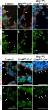
Tie2-Cre efficiently excises BRG1 and CHD4 in Brg1fl/fl; Chd4fl/fl:Cre+ endothelial cells. E10.5 embryos were cryosectioned and immunostained with an anti-BRG1 antibody (green) (A to F) or an anti-CHD4 antibody (green) (G to L) and an anti-PECAM antibody (red) to mark endothelial cells. Nuclei were stained with Hoechst dye (blue). Arrows designate individual endothelial cells. (A to F) Brg1fl/fl:Cre+ (B and E) and Brg1fl/fl; Chd4fl/fl:Cre+ (C and F) endothelial cells display significantly reduced expression of BRG1 compared to control cells (A and D). Likewise, Chd4fl/fl:Cre+ (H and K) and Brg1fl/fl; Chd4fl/fl:Cre+ (I and L) endothelial cells have considerably reduced expression of CHD4 compared to controls (G and J). (A to L) Scale bars, 50 μm.
Fig 3
Fig 3
Chd4 deletion does not rescue Brg1fl/fl:Cre+ anemia. (A to D) Gross photos of E10.5 control and mutant embryos with attached yolk sacs and placentae. Vessels in control (A) and Chd4fl/fl:Cre+ (C) yolk sacs contain visible red blood. Vessels in Brg1fl/fl:Cre+ (B) and Brg1fl/fl; Chd4fl/fl:Cre+ (D) yolk sacs are pale. Arrowheads designate blood vessels. (E to H) Cryosections from E10.5 control and mutant yolk sacs were stained by TUNEL (green) to identify apoptotic cells. Primitive erythrocytes in control (E) and Chd4fl/fl:Cre+ (G) yolk sac vessels are TUNEL negative. A subset of primitive erythrocytes in Brg1fl/fl:Cre+ (F) and Brg1fl/fl; Chd4fl/fl:Cre+ (H) yolk sac vessels are TUNEL positive (arrows). Nuclei were stained with Hoechst dye (blue). Yolk sac vessels are outlined (yellow). (I to L) Cryosections from E10.5 control and mutant embryos were stained with benzidine (yellow) for detection of hemoglobin in developing blood cells. The majority of primitive erythrocytes in control (I) and Chd4fl/fl:Cre+ (K) vessels stained with benzidine. In contrast, the majority of primitive erythrocytes in Brg1fl/fl:Cre+ (J) and Brg1fl/fl; Chd4fl/fl:Cre+ (L) vessels failed to stain with benzidine (arrows). Scale bars, 1 mm (A to D), 50 μm (E to H), and 100 μm (I to L).
Fig 4
Fig 4
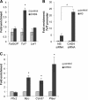
CHD4 acts downstream of β-catenin to modulate vascular Wnt signaling. (A to C) Endothelial cells from littermate control and mutant yolk sacs were isolated, RNA was purified, and cDNA was synthesized. qPCR was performed using a custom-designed array containing primer sets for 28 Wnt target genes. Data from at least four independent experiments were combined and analyzed using SABiosciences Excel-based software. Pie charts display the percentage of genes in each group that were downregulated, upregulated, or unchanged in Brg1fl/fl:Cre+ (A) or Chd4fl/fl:Cre+ (B) YSECs compared to littermate control YSECs. (C) A Venn diagram shows the transcripts that were downregulated in Brg1fl/fl:Cre+ YSECs compared to control YSECs and upregulated in Chd4fl/fl:Cre+ YSECs compared to control YSECs. (D to E) C166 yolk sac endothelial cells were transfected with nonspecific (NS) or CHD4-specific siRNA oligonucleotides for 48 h. (D) Western blot analysis was performed using antibodies that recognize CHD4, β-catenin, or GAPDH. β-Catenin band intensity was determined and normalized to the intensity of GAPDH. The results from five independent experiments were combined, and data are presented as the means ± SEM. (E) RNA was isolated, cDNA was synthesized, and qPCR was performed using gene-specific primers (Chd4, β-catenin, Tcf3, Tcf4, Tcf7, and Lef1). Relative fold change was calculated for transcripts in CHD4 knockdown cells and normalized to nonspecific siRNA-transfected samples (dotted line). Error bars represent ±SEM of results from four independent experiments. Significant differences were calculated using a two-tailed Student t test (*, P < 0.05).
Fig 5
Fig 5
CHD4 modulates vascular Wnt signaling at two levels. (A) Chromatin immunoprecipitation (ChIP) assays were performed using antibodies against CHD4 or a polyhistidine epitope tag as a negative control. DNA was isolated and amplified by qPCR to determine whether CHD4 bound the promoter region of Tcf7 or Lef1. (B) Chromatin harvested from nonspecific (NS) or CHD4 siRNA-transfected C166 yolk sac endothelial cells was immunoprecipitated with an antibody against histone H3 or a negative-control antibody. DNA was isolated and amplified by qPCR to examine the relative nucleosome density at the Tcf7 promoter. (C) Chromatin immunoprecipitation assays were carried out using antibodies against CHD4 or IgG (negative control) to determine whether CHD4 was associated with the promoter region of the Wnt target genes Pitx2, Myc, Ccnd1, and Plaur. (A to C) A region greater than 5 kb upstream of the Fzd5 transcription start site (Fzd5UP) was used as a negative control. Data from three independent experiments were combined and are presented as fold enrichment over the level with the negative-control antibody. Significant differences were calculated using a two-tailed Student t test (*, P < 0.05).
Fig 6
Fig 6
A subset of Wnt target genes are rescued in Brg1fl/fl; Chd4fl/fl:Cre+ YSECs and LiCl-treated Brg1fl/fl:Cre+ YSECs. Endothelial cells from littermate control and mutant yolk sacs were isolated, RNA was purified, and cDNA was synthesized. (A to D) qPCR was performed using a custom-designed array containing primer sets for 28 Wnt target genes. Data from at least four independent experiments were combined and analyzed using SABiosciences Excel-based software. (A to B) Pie charts display the percentage of genes in each group that were downregulated, upregulated, or unchanged in Brg1fl/fl; Chd4fl/fl:Cre+ (A) or LiCl-treated Brg1fl/fl:Cre+ (B) YSECs compared to littermate control YSECs. (C to D) Venn diagrams summarize the transcripts that were downregulated in Brg1fl/fl:Cre+ YSECs compared to control YSECs, upregulated in Chd4fl/fl:Cre+ YSECs compared to control YSECs, and unchanged (i.e., rescued) in Brg1fl/fl; Chd4fl/fl:Cre+ YSECs compared to control YSECs (C) or downregulated in Brg1fl/fl:Cre+ YSECs compared to control YSECs and unchanged (i.e., rescued) in LiCl-treated Brg1fl/fl:Cre+ YSECs compared to LiCl-treated control YSECs (D). (E) qPCR was performed using gene-specific primers for Pitx2. Relative fold change was calculated and normalized to littermate control samples (dotted line). Error bars represent ±SEM of results from six independent experiments. Significant differences between littermate control and mutant samples were calculated using a two-tailed Student t test (*, P < 0.05).
Fig 7
Fig 7
Model for how BRG1 and CHD4 antagonistically influence Wnt signaling during yolk sac vascular development. BRG1 activates the Wnt signaling pathway in two ways: through transcriptional regulation of multiple Fzd receptor genes upstream of β-catenin and through coregulation of Wnt target genes downstream of β-catenin (12). CHD4 represses the Wnt signaling pathway downstream of β-catenin through transcriptional regulation of the Wnt-responsive transcription factor Tcf7 and select Wnt target genes.

Similar articles

Cited by

References

    1. Bultman S, et al. 2000. A Brg1 null mutation in the mouse reveals functional differences among mammalian SWI/SNF complexes. Mol. Cell 6:1287–1295 - PubMed
    1. Bultman SJ, Gebuhr TC, Magnuson T. 2005. A Brg1 mutation that uncouples ATPase activity from chromatin remodeling reveals an essential role for SWI/SNF-related complexes in beta-globin expression and erythroid development. Genes Dev. 19:2849–2861 - PMC - PubMed
    1. Chappell JC, Bautch VL. 2010. Vascular development: genetic mechanisms and links to vascular disease. Curr. Top. Dev. Biol. 90:43–72 - PubMed
    1. Corada M, et al. 2010. The Wnt/beta-catenin pathway modulates vascular remodeling and specification by upregulating Dll4/Notch signaling. Dev. Cell 18:938–949 - PMC - PubMed
    1. Dejana E. 2010. The role of wnt signaling in physiological and pathological angiogenesis. Circ. Res. 107:943–952 - PubMed

Publication types

MeSH terms

LinkOut - more resources