. 2012 Apr;32(8):1542-54.
doi: 10.1128/MCB.05989-11. Epub 2012 Jan 30.

YY1 controls immunoglobulin class switch recombination and nuclear activation-induced deaminase levels

Affiliations

YY1 controls immunoglobulin class switch recombination and nuclear activation-induced deaminase levels

Kristina Zaprazna et al. Mol Cell Biol. 2012 Apr.

Abstract

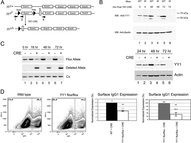
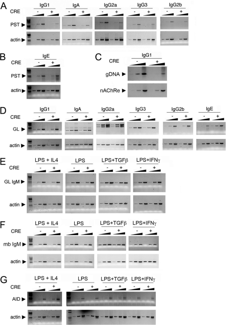
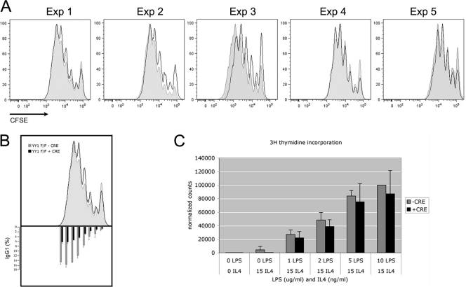
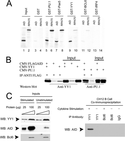
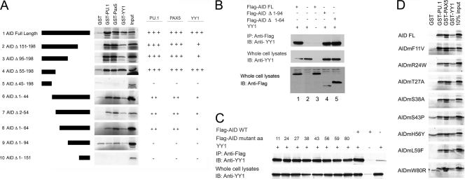
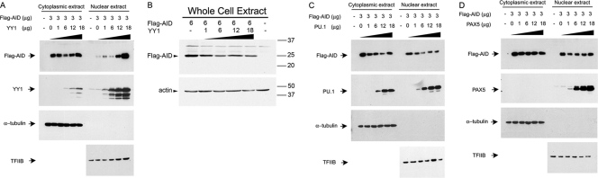
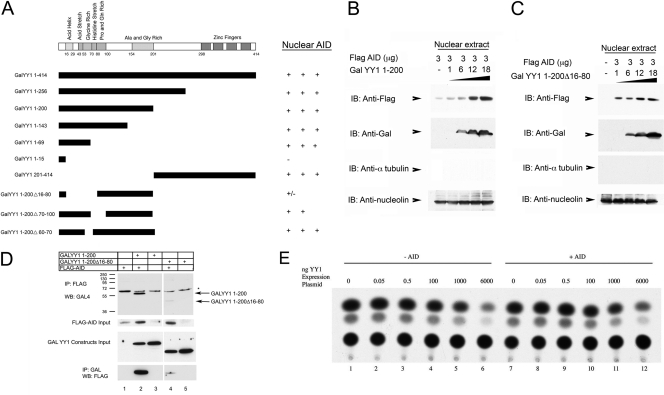
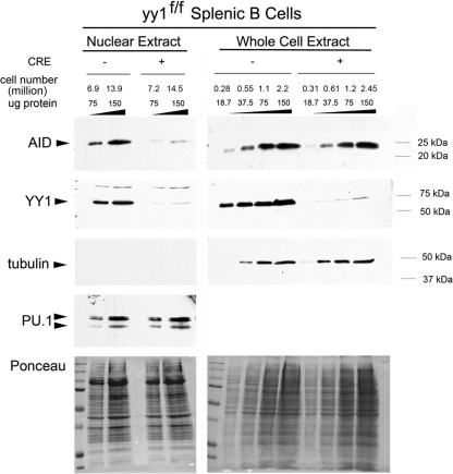
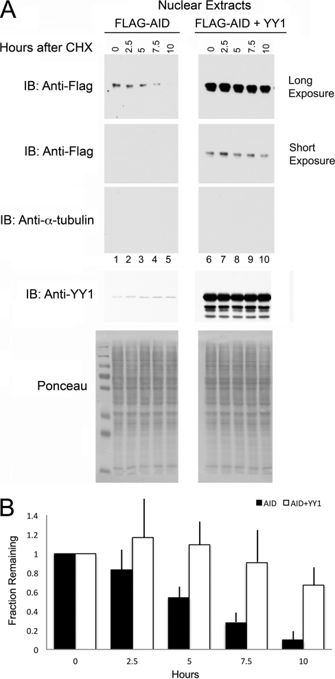
Activation-induced deaminase (AID) is an enzyme required for class switch recombination (CSR) and somatic hypermutation (SHM), processes that ensure antibody maturation and expression of different immunoglobulin isotypes. AID function is tightly regulated by tissue- and stage-specific expression, nuclear localization, and protein stability. Transcription factor YY1 is crucial for early B cell development, but its function at late B cell stages is unknown. Here, we show that YY1 conditional knockout in activated splenic B cells interferes with CSR. Knockout of YY1 did not affect B cell proliferation, transcription of the AID and IgM genes, or levels of various switch region germ line transcripts. However, we show that YY1 physically interacts with AID and controls the accumulation of nuclear AID, at least in part, by increasing nuclear AID stability. We show for the first time that YY1 plays a novel role in CSR and controls nuclear AID protein levels.


Figures

Fig 1
Fig 1
Conditional YY1 knockout reduces CSR and surface IgG1 expression in activated mouse B cells. (A) Schematic representation of YY1 loci from wild-type (yy1+), yy1 floxed (yy1f), and yy1 CRE-deleted (yy1Δ) mice. The arrows show the locations of primers that detect floxed (primers 1 and 2) and deletion (primers 1 and 4) alleles (30). (B) Recombinant TAT-CRE protein efficiently deletes the floxed yy1 gene in primary splenic B cells ex vivo. Splenic B cells were isolated from yy1flox/flox mice and then treated with recombinant TAT-CRE protein. Cells were harvested at 0, 48, or 72 h (top) or at 18, 48, and 72 h (bottom) after TAT-CRE addition, and whole-cell lysates were assayed for the presence of YY1 by Western blotting (WB) with anti-YY1 antibody. YY1 levels were greatly reduced at 48 h and nearly undetectable at 72 h. Western blotting with anti-β-actin antibody served as a loading control. (C) Genotyping of yy1flox/flox splenic B cells treated with TAT-CRE. Cells were harvested at 0, 18, 48, and 72 h after TAT-CRE addition. Genomic DNA was subjected to PCR with primers that detect the yy1 floxed allele or the yy1-deleted allele. Primers to the Cκ constant region served as loading controls. (D) Splenic B cells were purified from wild-type (WT) or yy1flox/flox mice and treated with recombinant TAT-CRE protein. Levels of surface IgG1 were determined by FACS (left). A histogram of surface IgG1 expression in wild-type or yy1flox/flox B cells treated with TAT-CRE is shown (second from right). Alternatively, yy1flox/flox B cells were treated with TAT-CRE or were mock treated with vehicle control (far right). After a 3-day induction with LPS and IL-4, surface IgG1 expression was measured by FACS. Dead cells were excluded from analysis based on 7-AAD staining. Levels of surface IgG1 expression are shown from 6 or 14 independent experiments (second from right and far right, respectively). The error bars indicate standard deviations, and the asterisks indicate significance (P < 0.001).
Fig 2
Fig 2
YY1 regulates CSR, but not germ line, IgM or Aicda transcripts. (A to G) Splenic B cells purified from yy1flox/flox mice were treated with bacterial TAT-CRE, resulting in YY1 knockout (CRE +), or were mock treated (CRE −). The cells were treated with LPS plus IL-4 to induce CSR to IgG1 and IgE, with LPS to induce CSR to IgG3 and IgG2b, with LPS plus TGF-β to induce CSR to IgA, and with LPS plus IFN-γ to induce CSR to IgG2a. RNA was isolated and assayed for PSTs (A and B) or germ line switch region transcripts (GL) (D) by reverse transcription (RT)-PCR. Fivefold-increasing sample amounts are shown for each isotype or loading control. The data are representative of three independent experiments. (A) Loss of YY1 reduces CSR to IgG1, IgA, IgG2a, IgG3, and IgG2b as assayed by RT-PCR of PSTs. (B) Loss of YY1 does not affect CSR to IgE. (C) Digestion circularization PCR of DNA from splenic B cells activated with LPS and IL-4 for 4 days. Primers that detect switching to IgG1 (genomic DNA [gDNA]) were used in the top gel, and primers for the loading control nicotinic acetylcholine receptor (nAChRe) β-subunit gene were used in the bottom gel. The amount of gDNA was increased by 5-fold in each lane. (D) Loss of YY1 has no impact on switch region germ line transcripts (GL), except for a 5-fold increase in IgE transcripts. (E and F) IgM germ line transcripts (GL IgM) and membrane-bound transcripts (mb IgM) are not impacted by loss of YY1. (G) AID mRNA expression is not impacted by loss of YY1.
Fig 3
Fig 3
Loss of YY1 does not affect proliferation of activated B cells. (A) Splenic B cells from yy1flox/flox mice were stained with CFSE and either untreated (shaded profiles) or treated with TAT-CRE (open profiles). After a 3-day stimulation with LPS and IL-4, CFSE levels in each cell were determined by FACS. The results of 5 independent experiments are shown. (B) Surface IgG1 expression in cells stained with CFSE in panel A was determined by FACS (bottom). Averages from 5 experiments are presented, normalized to surface IgG1 expression in division cycle number 5. The error bars indicate standard deviations. (C) [3H]thymidine incorporation of splenic B cells from yy1flox/flox mice cultured for 3 days in LPS and IL-4 media and treated with TAT-CRE (+CRE) or mock treated (−CRE). The averages of 3 experiments reflect counts normalized to mock-treated cells cultured in 10 μg/ml LPS and 15 ng/ml IL-4. The error bars indicate standard deviations.
Fig 4
Fig 4
AID interacts with PU.1, PAX5, and YY1 in vitro and in vivo. (A) GST pulldown assay. In vitro-translated 35S-labeled AID or RPA70 protein was incubated with GST-tagged PU.1, PAX5, YY1, Bcl6, or IRF4 purified from bacteria in the presence of ethidium bromide to inhibit protein-DNA interactions and evaluated by SDS-PAGE. (B) AID coimmunoprecipitates with YY1 and PU.1 in vivo. 293T cells were transfected with CMV–Flag-AID, CMV-YY1, or CMV-PU.1 plasmid in the combinations indicated. After immunoprecipitation (IP) with anti-Flag antibody, levels of YY1 and PU.1 were detected by Western blotting with anti-YY1 and anti-PU.1 antibodies. Two percent (10 μg) of the sample is shown as input. The data are representative of three independent experiments. (C) AID and YY1 interact in vivo in B cells. Cell lysates from either stimulated or unstimulated CH12 cells were assayed by Western blotting for YY1, AID, and BCL6 expression (left). The same lysates were immunoprecipitated with the antibodies shown (right), and AID was detected by Western blotting.
Fig 5
Fig 5
Identification of AID sequences necessary for interaction with YY1, PU.1, and PAX5. (A) GST pulldown assays were performed with GST-tagged YY1, PU.1, or PAX5 and in vitro-translated 35S-labeled AID deletion mutants. Specific AID deletion mutants are shown on the left, and levels of interaction with PU.1, PAX5, and YY1 are summarized on the right (+, about 25%; ++, about 50%; +++, 100%; −, not above background). (B) YY1 coimmunoprecipitates with Flag-AIDΔ1-94. 293T cells were transfected with the expression plasmids shown above each lane. Samples were immunoprecipitated with anti-Flag antibody and subjected to the Western blot (IB) procedure with anti-YY1 antibody. At the bottom are shown various Flag-AID and mutant inputs. FL, full length. (C) Various hyper-IgM AID point mutants coimmunoprecipitate with YY1. The Flag-AID wild-type and point mutants shown above the lanes were cotransfected into 293T cells with or without YY1 expression plasmid. Samples were immunoprecipitated with anti-Flag antibody and subjected to the Western blot procedure with anti-YY1 antibody. (D) Various hyper-IgM syndrome AID point mutants interact with YY1, PU.1, and Pax5. GST pulldown assays were performed with the 35S-labeled proteins shown on the left and GST fusion proteins shown above each lane.
Fig 6
Fig 6
YY1 affects AID nuclear localization in transfected 293T cells. (A to D) Flag-AID was transfected to 293T cells with increasing amounts of YY1 (A and B), PU.1 (C), or PAX5 (D) expression plasmids. Two days after transfection, nuclear, cytoplasmic, or whole-cell extracts were prepared and assayed by Western blotting for expression of AID with anti-Flag antibody, as well as for expression of YY1, PU.1, and PAX5 by Western blotting with appropriate antibodies. Sixty micrograms of protein assayed in each sample represents about 0.3 million cells of cytoplasmic extract and 3 million cells of nuclear extracts. The purity of nuclear extracts is shown by absence of cytoplasmic α-tubulin, and sample loading is controlled by levels of TFIIB. The data are representative of three independent experiments.
Fig 7
Fig 7
Mapping of YY1 domains needed for increased AID nuclear accumulation. (A) Various GALYY1 mutants (diagrammed on the left) were cotransfected into 293T cells with Flag-AID. Levels of nuclear AID accumulation are represented on the right. (B and C) GALYY1 1-200 or GALYY1 1-200Δ16-80 was cotransfected with Flag-AID into 293T cells. While GALYY1 1-200 expression led to increased AID nuclear accumulation (B), GALYY1 1-200Δ16-80 did not (C). (D) AID nuclear accumulation correlates with YY1 physical interaction. GALYY1 1-200 or GALYY1 1-200Δ16-80 was cotransfected with Flag-AID into 293T cells. Whole-cell extracts of the transfected cells immunoprecipitated with anti-Flag antibody and then blotted with anti-GAL antibody showed coprecipitation of GALYY1 1-200 with Flag-AID but not GALYY1 1-200Δ16-80 (top). Three percent input blots are shown in the middle gels. The lower gel shows the reciprocal experiment, in which immunoprecipitation was performed with anti-GAL antibody followed by blotting with anti-Flag antibody. Again, only GALYY1 1-200 interacted strongly with Flag-AID. (E) AID does not impact YY1 transcription functions. Increasing amounts of CMV-YY1 were transfected in the presence and absence of CMV–Flag-AID and assayed for transcriptional activity of a YY1-responsive promoter containing a multimerized YY1 binding site adjacent to the TK promoter, (YY1)4TKCAT. This promoter is activated by low levels of YY1 but repressed by high levels (6, 42). 293T cells were transfected with (YY1)4TKCAT reporter plasmid and increasing amounts of YY1 expression plasmid either with (+) or without (−) AID expression plasmid. The levels of YY1 transcriptional activation and repression were analyzed by chloramphenicol acetyltransferase assay.
Fig 8
Fig 8
YY1 regulates AID levels in nuclear extracts from activated mouse splenic B cells. Splenic B cells from yy1flox/flox mice were treated with bacterial TAT-CRE (+CRE) or mock-treated (−CRE) as a control and activated ex vivo with LPS plus IL-4. Nuclear and whole-cell extracts were made after 3 days in culture. Endogenous AID protein levels were determined by Western blotting with an anti-AID antibody (Cell Signaling). AID signals were normalized to tubulin and PU.1 levels, as well as the signal from reversible staining of Western blot membranes with Ponceau S solution. A representative of 3 independent experiments is shown. The representative number of cell equivalents for each lane is shown to indicate the much larger cell numbers needed for the nuclear extract lanes compared to whole-cell extracts.
Fig 9
Fig 9
YY1 controls AID stability in the nucleus. (A) Flag-AID or Flag-AID plus YY1 was cotransfected into 293T cells, and 2 days later, the cells were treated with 100 μg/ml cycloheximide (CHX) to inhibit protein synthesis. Cells were harvested at the times indicated above the lanes, and nuclear extracts were immunoblotted with antibodies to Flag (top two gels; long and shorter exposures, respectively), α-tubulin to detect potential cytoplasmic contamination (third gel from top), and YY1 (fourth gel from top). Comparable loading of each lane is shown by Ponceau staining in the bottom gel. The exposures of the top two gels were adjusted to enable comparison of the stability of AID alone (top gel, lanes 1 to 5) with that of AID plus YY1 (second gel from top, lanes 6 to 10). (B) The fractions of AID remaining normalized to nucleolin and Ponceau S staining are shown for AID versus AID plus YY1 for 8 independent experiments. The error bars show the standard deviations of the mean.

References

    1. Aoufouchi S, et al. 2008. Proteasomal degradation restricts the nuclear lifespan of AID. J. Exp. Med. 205:1357–1368 - PMC - PubMed
    1. Atchison L, Ghias A, Wilkinson F, Bonini N, Atchison ML. 2003. The YY1 transcription factor functions as a PcG protein in vivo. EMBO J. 22:1347–1358 - PMC - PubMed
    1. Ballantyne J, Henry DL, Marcu KB. 1997. Antibody class switch recombinase activity is B cell stage specific and functions stochastically in the absence of ‘targeted accessibility’ control. Int. Immunol. 9:963–974 - PubMed
    1. Basu U, et al. 2011. The RNA exosome targets the AID cytidine deaminase to both strands of transcribed duplex DNA substrates. Cell 144:353–363 - PMC - PubMed
    1. Brar SS, Watson M, Diaz M. 2004. Activation-induced cytosine deaminase (AID) is actively exported out of the nucleus but retained by the induction of DNA breaks. J. Biol. Chem. 279:26395–26401 - PubMed

Publication types

Substances

LinkOut - more resources