

Cerebral perfusion changes in migraineurs: a voxelwise comparison of interictal dynamic susceptibility contrast MRI measurements
- PMID: 22290556
- PMCID: PMC3743424
- DOI: 10.1177/0333102411435985
Cerebral perfusion changes in migraineurs: a voxelwise comparison of interictal dynamic susceptibility contrast MRI measurements
Abstract
Introduction: The increased risk of cerebro- and cardiovascular disease in migraineurs may be the consequence of a systemic condition affecting whole body vasculature. At cerebrovascular level, this may be reflected by interictal global or regional cerebral perfusion abnormalities. Whether focal perfusion changes occur during interictal migraine has not been convincingly demonstrated.
Methods: We measured brain perfusion with dynamic susceptibility contrast magnetic resonance imaging (DSC-MRI) in 29 interictal female migraineurs (12 migraine with aura (MA), 17 migraine without aura (MO)), and 16 female controls. Perfusion maps were compared between these groups with a voxelwise (p < 0.001, uncorrected, minimum cluster size 20 voxels) and a region-of-interest approach.
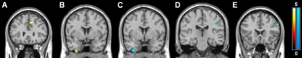
Results: In whole brain voxelwise analyses interictal hyperperfusion was observed in the left medial frontal gyrus in migraineurs and in the inferior and middle temporal gyrus in MO patients, in comparison with controls. Hypoperfusion was seen in the postcentral gyrus and in the inferior temporal gyrus in MA patients and in the inferior frontal gyrus in MO patients. Additional focal sites of hyperperfusion were noted in subgroups based on attack frequency and disease history. Region-of-interest analyses of the pons, hypothalamus, occipital lobe, and cerebellum did not show interictal perfusion differences between migraineurs and controls.
Conclusions: We conclude that interictal migraine is characterized by discrete areas of hyper- and hypoperfusion unspecific for migraine pathophysiology and not explaining the increased vulnerability of particular brain regions for cerebrovascular damage.
Conflict of interest statement
Ona Wu has a patent on “Delay-compensated calculation of tissue blood flow,” US Patent 7,512,435. March 31, 2009, and the patent has been licensed to General E, Siemens, and Olea Medical. The remaining authors declare that there is no conflict of interest regarding the subject matter of this article.
Figures

References
-
- Headache Classification Subcommittee of the International Headache Society. The International Classification of Headache Disorders. Cephalalgia. (2nd edition) 2004;24(Suppl 1):9–160. - PubMed
-
- Kruit MC, van Buchem MA, Hofman PA, Bakkers JT, Terwindt GM, Ferrari MD, et al. Migraine as a risk factor for subclinical brain lesions. JAMA. 2004;291:427–434. - PubMed
-
- Kruit MC, Launer LJ, Ferrari MD, van Buchem MA. Infarcts in the posterior circulation territory in migraine. The population-based MRI CAMERA study. Brain. 2005;128:2068–2077. - PubMed
Publication types
MeSH terms
Grants and funding
LinkOut - more resources
Full Text Sources
Medical
Miscellaneous
