. 2012 May;30(5):865-75.
doi: 10.1002/stem.1049.

SFRP1 and SFRP2 dose-dependently regulate midbrain dopamine neuron development in vivo and in embryonic stem cells

Affiliations

SFRP1 and SFRP2 dose-dependently regulate midbrain dopamine neuron development in vivo and in embryonic stem cells

Julianna Kele et al. Stem Cells. 2012 May.

Abstract

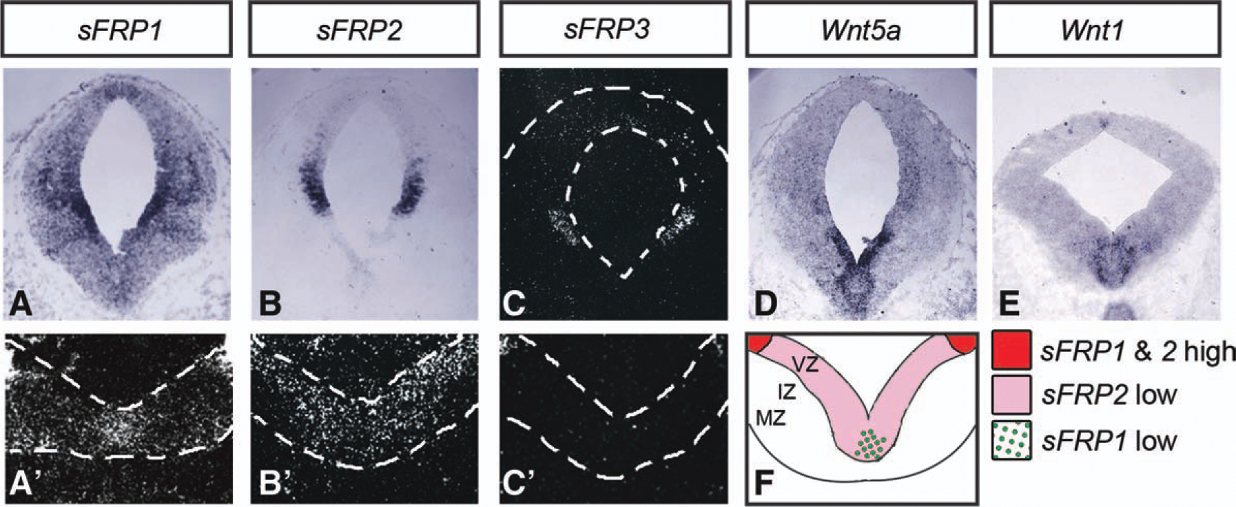
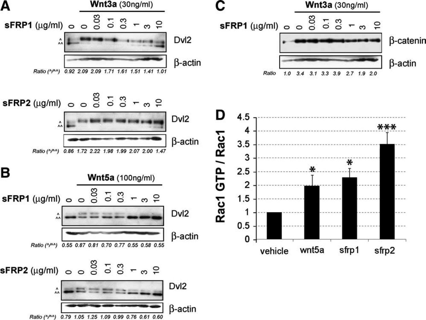
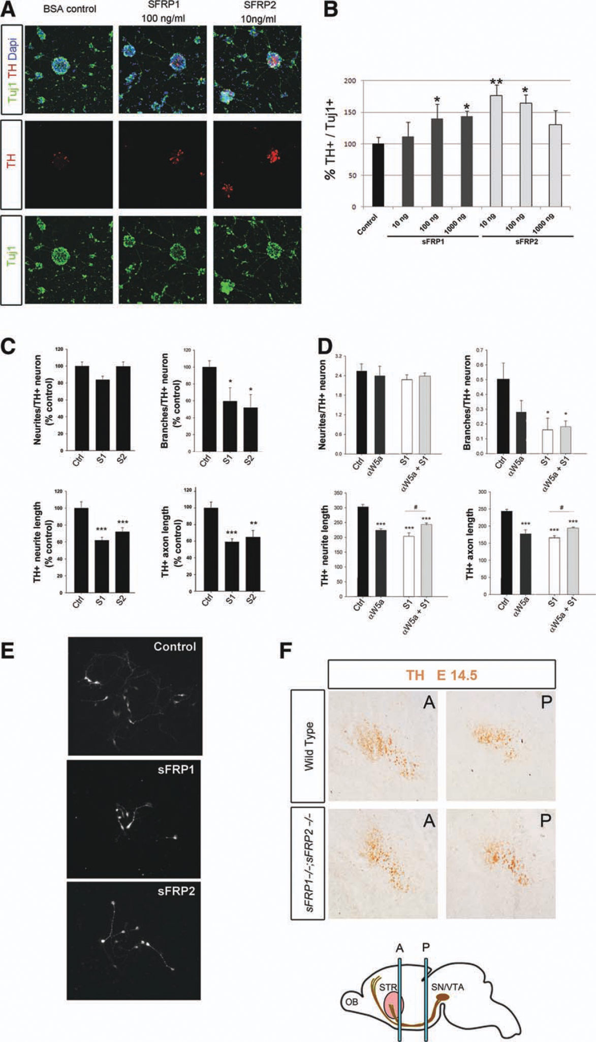
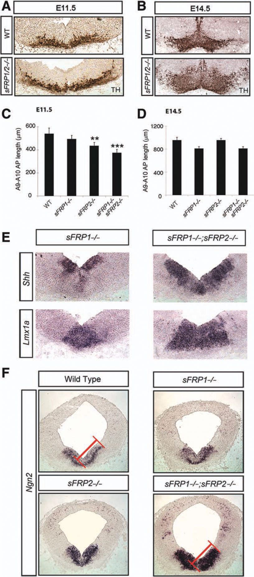
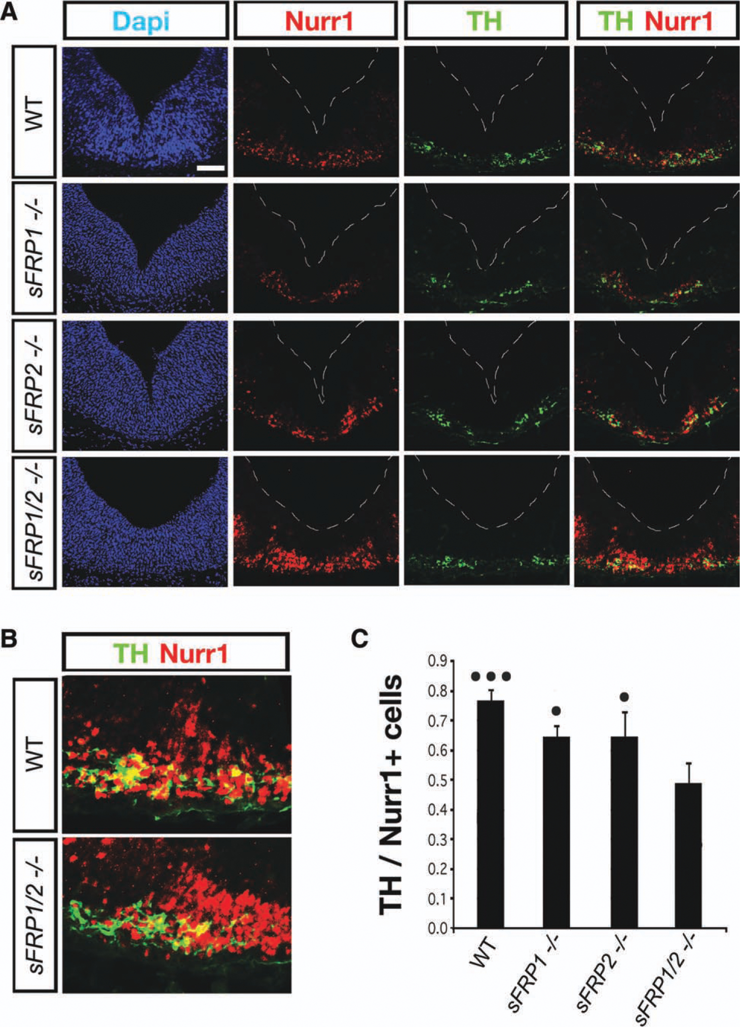
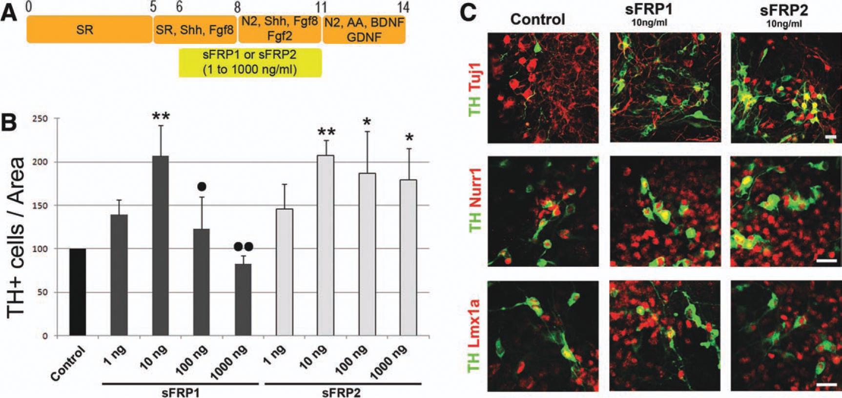
Secreted Frizzled related proteins (sFRPs) are a family of proteins that modulate Wnt signaling, which in turn regulates multiple aspects of ventral midbrain (VM) and dopamine (DA) neuron development. However, it is not known which Wnt signaling branch and what aspects of midbrain DA neuron development are regulated by sFRPs. Here, we show that sFRP1 and sFRP2 activate the Wnt/planar-cell-polarity/Rac1 pathway in DA cells. In the developing VM, sFRP1 and sFRP2 are expressed at low levels, and sFRP1-/- or sFRP2-/- mice had no detectable phenotype. However, compound sFRP1-/-;sFRP2-/- mutants revealed a Wnt/PCP phenotype similar to that previously described for Wnt5a-/- mice. This included an anteroposterior shortening of the VM, a lateral expansion of the Shh domain and DA lineage markers (Lmx1a and Th), as well as an accumulation of Nurr1+ precursors in the VM. In vitro experiments showed that, while very high concentrations of SFRP1 had a negative effect on cell survival, low/medium concentrations of sFRP1 or sFRP2 promoted the DA differentiation of progenitors derived from primary VM cultures or mouse embryonic stem cells (ESCs), mimicking the effects of Wnt5a. We thus conclude that the main function of sFRP1 and sFRP2 is to enhance Wnt/PCP signaling in DA cells and to regulate Wnt/PCP-dependent functions in midbrain development. Moreover, we suggest that low-medium concentrations of sFRPs may be used to enhance the DA differentiation of ESCs and improve their therapeutic application.


Conflict of interest statement

Disclosure of Potential Conflicts of Interest

The authors indicate no potential conflicts of interest.

Figures

Figure 1.
Figure 1.
Spatial pattern of expression of sFRP1, sFRP2, and sFRP3 in the developing midbrain at E11.5. (A–C, A′–C′): In situ hybridization for sFRP1, sFRP2, and sFRP3 mRNA on coronal sections of mouse midbrain at E11.5 revealed high expression in the midbrain AP. Higher magnification of radioactive in situ hybridization detected expression of sFRP1 in the midline and a low expression of sFRP2 in the whole ventral midbrain (VM). In contrast, low levels of sFRP3 were only detected laterally, in the midbrain AP but not in the VM. (D, E): Wnt5a and Wntl expression was also detected in the VM. (F): Scheme summarizing the overlap and expression levels of sFRP1 and sFRP2 in the VM. Abbreviations: sFRP, secreted Frizzled related protein; IZ, intermediate zone; MZ, marginal zone; VZ, ventricular zone.
Figure 2.
Figure 2.
SFRP1 and sFRP2 inhibit the Wnt3a- or Wnt5a-induced Dvl shift and activate Rac1 in a dopaminergic neuron cell line. SN4741 cells were treated with 30 ng/ml Wnt3a (A) or 100 ng/ml Wnt5a (B) and increasing concentrations (0, 0.03, 0.1, 0.3, 1, 3, and 10 μg/ml) of sFRP1or sFRP2. (A): The Wnt3a-induced Dvl2 phosphorylation mobility shift was reduced by a lower concentration of sFRP1 than sFRP2. (B): The Wnt5a-induced Dvl2 phosphorylation mobility shift was efficiently reduced by both sFRP1 and sFRP2. The ratio between the hyperphosphory-lated form (upper band ^) and the nonshifted lower band (^^) is given for representative gels (n ≥ 3). (C): SFRP1 also reduced the increase in active β-catenin induced by Wnt3a. The induction of active β-catenin was expressed as the ratio between the densitometric quantification of active β-catenin and the loading control actin (ratio: β-catenin/actin). (D): Both sFRP1 and sFRP2 (2 μg/ml) increased Rac1 activity by twofold and 3.5-fold, respectively, compared with vehicle. ***, p < .001 and *, p < .05 by one-way analysis of variance, with Turkey’s post hoc test for multiple comparisons (n = 3). Abbreviation: sFRP, secreted Frizzled related protein.
Figure 3.
Figure 3.
Differential effects of sFRP1 and sFRP2 on TH+ cell numbers and neuritogenesis. Mouse E11.5 ventral midbrain cultures were treated for 3 days with vehicle (BSA Ctrl) or different concentrations of sFRP1 or sFRP2 (10, 100, or 1,000 ng/ml) and analyzed for expression of beta III-tubulin (TuJ1, green) and TH (red), using DAPI (blue) as counterstaining. (A): SFRP1 (100 ng/ml) and sFRP2 (10 ng/ml) increased the percentage of Th+/Tuj1 + cells in primary cultures. (B): The percentage of TH+/TuJ1+ neurons increased in a dose-dependent manner in sFRP1- and sFRP2-treated primary cultures. Significant effects were detected at 100–1,000 ng/ml of sFRP1 and 10–100 ng/ml of sFRP2 as assessed by one-way analysis of variance with Tukey’s post hoc test for multiple comparisons (n = 3). p < .001; **, p < .01; *, p < .05. (C): The effects of sFRP1 or sFRP2 on neuritogenesis were evaluated in rat E13.5 primary precursor cells at concentrations capable of activating Rac1 (2.5 μg/ml, S1 or S2). DA neurite length was expressed in four different ways, as neurites/TH+ neuron, branches/TH+ neuron, length of the dominant neurite (axon), and average length per neurite. (D): Wnt5a effect was analyzed by adding a Wnt5a blocking antibody (αWnt5a) to the cultures, in combination with sFRP1 (S1). αWnt5a alone reduced the number of branches, TH+ neurite length, and TH+ axon length. Moreover, aWnt5a partially prevented the effect of sFRP1. ***, p < .001; **, p < .01; *, p < .05 by one-way analysis of varaiance, with Tukeys’s post hoc test for multiple comparisons (n = 3). (E): TH immunocytochemistry showing the reduction of neurite branching, length, and axonal length in SFRP1- and sFRP2-treated cultures. (F): TH im-munostaining showing DA axons at anterior and posterior levels of the medial forebrain bundle at E14.5 (A, P, STR [pink], SN/VTA [brown], and OB). Abbreviations: A, anterior level; bovine serum albumin, BSA; 4,6-diamidino-2-, DAP; OB, olfactory bulb; P, posterior level; STR, striatum; SN/VTA, substantia nigra and ventral tegmental area; sFRP, secreted Frizzled related protein; TH, tyrosine hydroxylase.
Figure 4.
Figure 4.
Convergent extension defects in the ventral midbrain of sFRP1−/−;sFRP2−/− mice. While the number of TH+ cells in the ventral midbrain (VM) was not altered in sFRP1−/−;sFRP2−/− mice, their medial-lateral distribution at E11.5 (A) and their medial-lateral and dorsal-ventral distribution at E14.5 (B) were increased. This was accompanied by a reduction in the rostral-caudal distribution of TH+ cells at E11.5 (C) but not at E14.5 (D). *** p < .001; **, p < .01 by one-way analyses of variance, with Bonferroni’s post hoc test for multiple comparisons (n = 4 for E11.5 and n = 3 for E14.5). In addition to the increased lateral distribution of TH, other markers such as Shh (floorplate [FP] and basal plate [BP]) and Lmxla (FP) were also laterally expanded in sFRP1−/−;sFRP2−/− mice at E11.5 (E). The expression of Ngn2 in the VM expanded dorsoven-trally, but not laterally, in sFRP1−/−;sFRP2−/− mice at E11.5 (F). Abbreviations: Ngn2, neurogenin 2; sFRP, secreted Frizzled related protein; TH, tyrosine hydroxylase; WT, wild type. Shh, Sonic hedgehog homolog; Lmx1a, LIM homeobox transcription factor 1 alpha.
Figure 5.
Figure 5.
Deletion of sFRP1, sFRP2, and sFRP1/2 reduced the differentiation of Nurr1+/TH− DA precursors into Nurr1+/TH+ DA neurons. Immunohistochemistry for Nurr1 revealed an increase in the medial-lateral distribution of Nurr1+ cells in sFRP1−/−;sFRP2−/− compared with WT (A). This increase in Nurr1+ cells did not result in an increase in the number of Nurr1+/TH+ neurons in sFRP1−/−;sFRP2−/− but rather in an accumulation of Nurr1+/TH− DA precursors compared with WT (B). The proportion of TH+/Nurr1+ cells decreased modestly in sFRP1−/− or sFRP2−/− mice (n = 4 and 5, respectively). sFRP1−/−;sFRP2−/− mice (n = 5) showed a greater reduction in the proportion of TH+/Nurr1+ cells (C), which was significantly different from single mutants (•p < .05) and WT (•••p < .001), as assessed by analysis of variance with Tukey’s post hoc test. Our results thus suggest that the effects of endogenous low levels of sFRP1 and sFRP2 are additive. Abbreviations: DAPI, 4′,6-diami-dino-2-phenylindole; sFRP, secreted Frizzled related protein; TH, tyrosine hydroxylase; WT, wild type. Nurr1, nuclear receptor related 1.
Figure 6.
Figure 6.
SFRP1 and sFRP2 promote the generation of dopamine (DA) neurons from mouse embryonic stem cells (mESCs). (A): Schematic diagram illustrating the culture conditions used to generate DA neurons from mESCs. mESCs were cultured in the presence of serum replacement medium (SRM), followed by basal conditions, including SRM with FGF8 (for 3 days), SRM with FGF2 and FGF8 (for 3 days), and finally N2 supplement with AA, GDNF, and BDNF (for 3 days). Modified treatment conditions include the addition of sFRP1 or sFRP2 from day 6 to day 11 (from 1 to 1,000 ng/ml). (B): SFRP1 and sFRP2 increased the numbers of TH+ cells per Tuj1+ area up to a concentration of 10 ng/ml. While higher concentrations of sFRP1 decreased the numbers of TH, the effects of sFRP2 were sustained. *, p < .05; **, p < .001 compared with control; •, p < .05; ••, p < .01 compared with 10 ng/ml sFRP1, by one-way analysis of variance, with Tukey’s post hoc test for multiple comparisons. (C): SFRP1 or sFRP2 (10 ng/ml) did not affect Tuj1 expression but increased the expression of Nurr1 and Lmx1a in TH+ cells. The data show representative results from five independent differentiation experiments. Abbreviations: AA, ascorbic acid; BDNF, brain-derived neurotrophic factor; FGF, fibroblast growth factor; GDNF, glial-derived neurotrophic factor; sFRP, secreted Frizzled related protein; TH, tyrosine hydroxylase.

References

    1. Rattner A, Hsieh JC, Smallwood PM et al. A family of secreted proteins contains homology to the cysteine-rich ligand-binding domain of frizzled receptors. Proc Natl Acad Sci USA 1997;94: 2859–2863. - PMC - PubMed
    1. Lopez-Rios J, Esteve P, Ruiz JM et al. The Netrin-related domain of Sfrp1 interacts with Wnt ligands and antagonizes their activity in the anterior neural plate. Neural Dev 2008;3:19. - PMC - PubMed
    1. Uren A, Reichsman F, Anest V et al. Secreted frizzled-related protein-1 binds directly to Wingless and is a biphasic modulator of Wnt signaling. J Biol Chem 2000;275:4374–4382. - PubMed
    1. van Amerongen R, Nusse R. Towards an integrated view of Wnt signaling in development. Development 2009;136:3205–3214. - PubMed
    1. Ciani L, Salinas PC. WNTs in the vertebrate nervous system: From patterning to neuronal connectivity. Nat Rev Neurosci 2005;6: 351–362. - PubMed

Publication types

MeSH terms