. 2012 Mar 23;287(13):9672-9681.
doi: 10.1074/jbc.M111.328203. Epub 2012 Jan 30.

Proteome of Hydra nematocyst

Affiliations

Proteome of Hydra nematocyst

Prakash G Balasubramanian et al. J Biol Chem. .

Abstract

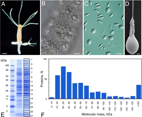
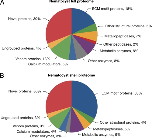
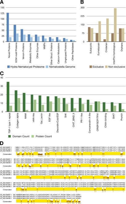
Stinging cells or nematocytes of jellyfish and other cnidarians represent one of the most poisonous and sophisticated cellular inventions in animal evolution. This ancient cell type is unique in containing a giant secretory vesicle derived from the Golgi apparatus. The organelle structure within the vesicle comprises an elastically stretched capsule (nematocyst) to which a long tubule is attached. During exocytosis, the barbed part of the tubule is accelerated with >5 million g in <700 ns, enabling a harpoon-like discharge (Nüchter, T., Benoit, M., Engel, U., Ozbek, S., and Holstein, T. W. (2006) Curr. Biol. 16, R316-R318). Hitherto, the molecular components responsible for the organelle's biomechanical properties were largely unknown. Here, we describe the proteome of nematocysts from the freshwater polyp Hydra magnipapillata. Our analysis revealed an unexpectedly complex secretome of 410 proteins with venomous and lytic but also adhesive or fibrous properties. In particular, the insoluble fraction of the nematocyst represents a functional extracellular matrix structure of collagenous and elastic nature. This finding suggests an evolutionary scenario in which exocytic vesicles harboring a venomous secretome assembled a sophisticated predatory structure from extracellular matrix motif proteins.


Figures

FIGURE 1.
FIGURE 1.
Nematocyst morphology and protein spectrum. A, H. magnipapillata. B, close-up of undischarged nematocysts assembled in battery cells of the tentacles. C, light microscopic view of isolated nematocyst capsules. D, scanning electron microscopic image of isolated nematocyst. E, one-dimensional SDS-PAGE of a nematocyst sample. The gel slices used for mass spectrometry are indicated. F, molecular mass distribution of the nematocyst proteome. Scale bars = 0.5 mm (A), 15 μm (B and C), and 2 μm (D).
FIGURE 2.
FIGURE 2.
Distribution of functional nematocyst proteome. A, functional protein classes of the total Hydra nematocyst proteome. B, functional protein classes of the reduced proteome from nematocyst shells generated by extensive SDS washing.
FIGURE 3.
FIGURE 3.
A, homologs of Hydra nematocyst proteins identified in the Nematostella genome. MMPs, matrix metalloproteases. B, evolutionary distribution of the 410 nematocyst proteins. Nematocyst proteins have been assigned to categories based on the presence of significant BLAST hits and Pfam domains. The “exclusive” distribution refers to a hierarchical classification in which each protein was assigned to only one category. Proteins with significant BLAST hits in basal eukaryotes (T. adhaerens or M. brevicollis) have been classified as “eukaryotic.” The remaining proteins with significant similarity in H. sapiens, D. melanogaster, Danio rerio, C. elegans, or B. floridae were binned as “eumetazoan.” Proteins that were not classified as either eukaryotic or eumetazoan but with significant hits in the N. vectensis genome were classified as “cnidarian.” The remaining proteins were further classified as “InterPro-homologous” if they had a significant hit against the InterPro Database or as “orphans” otherwise. For the “non-exclusive” distribution, each nematocyst protein was assigned to every category where a significant hit could be identified. C, domain distribution of Hydra nematocyst proteins with a significant Pfam hit but no significant BLAST hit to eukaryotic, eumetazoan, or other cnidarian proteomes. Dark green, total number of occurrences of a domain in the data set; light green, number of proteins in which a domain occurs. vWA, von Willebrand factor A; SCP, sperm coat protein. D, alignment of the proteins corresponding to the second largest cluster of orphan proteins (see “Experimental Procedures”). The conserved region is predicted to be globular and thus likely to represent a previously uncharacterized protein domain. MAM, meprin, A-5 protein, receptor protein tyrosine phosphatase mu; Cub, complement C1r/C1s, Uegf, bone morphogenetic protein; ShkT, Stichodactyla toxin domain; Shk, Stichodactyla-like domain.
FIGURE 4.
FIGURE 4.
Domain organization of ECM-type nematocyst proteins. PP, polyproline domain; SCP, sperm coat protein-like extracellular domain; vWFA, von Willebrand factor A domain. Gly-X-Y is the collagen triple helix.
FIGURE 5.
FIGURE 5.
Primary sequence and expression pattern of Cnidoin. A, repetitive motif in the glycine-rich elastic domain of Cnidoin. The central consensus sequence showing homology to spidroin-2 is boxed. B–D, whole mount double immunostaining with anti-Cnidoin (red) and anti-minicollagen-1 (green) antibodies. D demonstrates the co-localization of the two signals in the developing capsule walls of nematocysts. Scale bar = 10 μm. E, light microscopic image of fibers formed by purified recombinant Cnidoin in urea. Scale bar = 30 μm.
FIGURE 6.
FIGURE 6.
Schematic representation of hypothetic evolution of cnidarian nematocyst and ECM assembly. The secretome of early heterotrophic eukaryotes exhibited secretory vesicles containing enzymes used for extracellular digestion. On the basis of our findings (Fig. 4), we presume that, in the secretome, structural proteins evolved that finally gave rise to a variety of protozoan extrusive organelles exhibiting a variety of highly sophisticated discharge mechanisms. Some of these extrusomes exhibit a surprising structural similarity to nematocysts, suggesting that nematocysts may have evolved from extrusomes of a non-cnidarian precursor.

References

    1. Nüchter T., Benoit M., Engel U., Ozbek S., Holstein T. W. (2006) Nanosecond-scale kinetics of nematocyst discharge. Curr. Biol. 16, R316–R318 - PubMed
    1. Fry B. G., Wroe S., Teeuwisse W., van Osch M. J., Moreno K., Ingle J., McHenry C., Ferrara T., Clausen P., Scheib H., Winter K. L., Greisman L., Roelants K., van der Weerd L., Clemente C. J., Giannakis E., Hodgson W. C., Luz S., Martelli P., Krishnasamy K., Kochva E., Kwok H. F., Scanlon D., Karas J., Citron D. M., Goldstein E. J., McNaughtan J. E., Norman J. A. (2009) A central role for venom in predation by Varanus komodoensis (Komodo dragon) and the extinct giant Varanus (Megalania) priscus. Proc. Natl. Acad. Sci. U.S.A. 106, 8969–8974 - PMC - PubMed
    1. Fry B. G., Roelants K., Champagne D. E., Scheib H., Tyndall J. D., King G. F., Nevalainen T. J., Norman J. A., Lewis R. J., Norton R. S., Renjifo C., de la Vega R. C. (2009) The toxicogenomic multiverse: convergent recruitment of proteins into animal venoms. Annu. Rev. Genomics Hum. Genet. 10, 483–511 - PubMed
    1. Eble J. A. (2010) Matrix biology meets toxinology. Matrix Biol. 29, 239–247 - PubMed
    1. Mariscal R. N. (1974) Scanning electron microscopy of the sensory surface of the tentacles of sea anemones and corals. Z. Zellforsch. 147, 149–156 - PubMed

Publication types

LinkOut - more resources