. 2012 Jan;8(1):e1002500.
doi: 10.1371/journal.ppat.1002500. Epub 2012 Jan 26.

The bacterial cytoskeleton modulates motility, type 3 secretion, and colonization in Salmonella

Affiliations

The bacterial cytoskeleton modulates motility, type 3 secretion, and colonization in Salmonella

David M Bulmer et al. PLoS Pathog. 2012 Jan.

Abstract

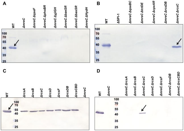
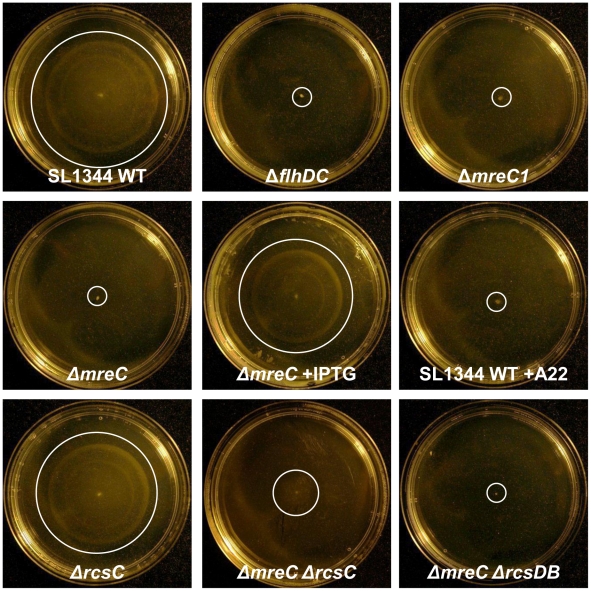
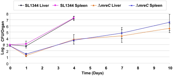
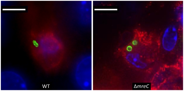
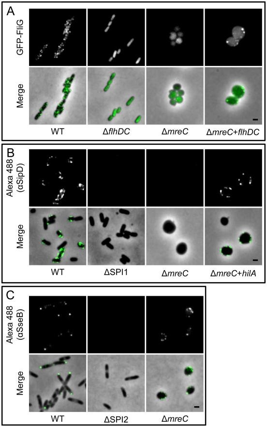
Although there have been great advances in our understanding of the bacterial cytoskeleton, major gaps remain in our knowledge of its importance to virulence. In this study we have explored the contribution of the bacterial cytoskeleton to the ability of Salmonella to express and assemble virulence factors and cause disease. The bacterial actin-like protein MreB polymerises into helical filaments and interacts with other cytoskeletal elements including MreC to control cell-shape. As mreB appears to be an essential gene, we have constructed a viable ΔmreC depletion mutant in Salmonella. Using a broad range of independent biochemical, fluorescence and phenotypic screens we provide evidence that the Salmonella pathogenicity island-1 type three secretion system (SPI1-T3SS) and flagella systems are down-regulated in the absence of MreC. In contrast the SPI-2 T3SS appears to remain functional. The phenotypes have been further validated using a chemical genetic approach to disrupt the functionality of MreB. Although the fitness of ΔmreC is reduced in vivo, we observed that this defect does not completely abrogate the ability of Salmonella to cause disease systemically. By forcing on expression of flagella and SPI-1 T3SS in trans with the master regulators FlhDC and HilA, it is clear that the cytoskeleton is dispensable for the assembly of these structures but essential for their expression. As two-component systems are involved in sensing and adapting to environmental and cell surface signals, we have constructed and screened a panel of such mutants and identified the sensor kinase RcsC as a key phenotypic regulator in ΔmreC. Further genetic analysis revealed the importance of the Rcs two-component system in modulating the expression of these virulence factors. Collectively, these results suggest that expression of virulence genes might be directly coordinated with cytoskeletal integrity, and this regulation is mediated by the two-component system sensor kinase RcsC.


Conflict of interest statement

The authors have declared that no competing interests exist.

Figures

Figure 1
Figure 1. Localization and morphological role of the S. Typhimurium Mre proteins.
(A) Fluorescence microscopy montage showing z-sections taken of MreB-GFP fusions in WT SL1344 revealing a helical distribution. Slices taken at 0.1 µm intervals on live cells in mid log phase going from left to right followed by maximum intensity projection (boxed). (B) Morphology of WT S. Typhimurium (I) and ΔmreC (II) reveal the mutant has changed from rod to round-shaped, with some heterogeneity in size noted. In all images the bar represents 1 µm.
Figure 2
Figure 2. Impact of ΔmreC on the transcription of flagellar genes.
Transcriptional expression profiles of flhD, flgA, fliA and fliC promoter reporters in WT SL1344 (blue diamonds) and ΔmreC (red squares) expressing the Photorhabdus luminescens LuxCDABE luciferase. Experiments were repeated at least three times and error bars indicate standard deviation.
Figure 3
Figure 3. Expression of Salmonella SPI-1 and SPI-2 effector proteins in ΔmreC.
(A) Expression of SPI-1 proteins in WT SL1344, ΔmreC, ΔSPI-1, and ΔSPI-2 mutants during SPI-1 inducing conditions as revealed by western blotting with polyclonal αSipA and αSipC antibodies. Expression of SPI-2 in WT SL1344, ΔmreC, ΔSPI-1, and ΔSPI-2 mutants during SPI-2 inducing conditions as revealed by western blotting of membrane fraction samples with polyclonal αSseB antibody. Samples representing total proteins and secreted proteins are shown. Arrows indicate the respective protein bands. (B)Transcriptional expression profiles of hilA, hilC, hilD, sopB (SPI-1) and ssaG (SPI-2) promoter reporters in WT SL1344 (blue diamonds) and ΔmreC (red squares). Experiments were repeated at least three times and error bars indicate standard deviation.
Figure 4
Figure 4. Percentage change in transepithelial resistance of differentiated Caco-2 cells after 4hr infection.
TER of polarised Caco-2 monolayers exposed to Salmonella strains at an MOI of 20. TER change is expressed as a percentage alteration at 4hr compared to the initial value at time zero. Error bars indicate the standard deviations derived from at least three independent experiments. * Indicates statistical difference from WT (p<0.05).
Figure 5
Figure 5. Western blotting screen of ΔmreC two-component system double mutants for recovery of SPI-1 T3S.
Panels A and B show western blots of total protein samples obtained from SL1344 WT, ΔSPI-1, ΔmreC, ΔmreC ΔqseF, ΔmreC ΔphoBR, ΔmreC ΔyjiGH, ΔmreC ΔbaeSR, ΔmreC ΔbasSR, ΔmreC ΔhydH, ΔmreC ΔqseBC, ΔmreC ΔtctDE, ΔmreC ΔcpxAR, ΔmreC ΔrcsDB, and ΔmreC ΔrcsC strains with αSipC antibody. Panels C and D show western blot of total protein samples obtained from SL1344 WT, ΔrcsA, ΔrcsB, ΔrcsC, ΔrcsD, ΔrcsF, ΔrcsDB, ΔrcsCBD and ΔmreC strains along with the ΔmreC ΔrcsA, ΔmreC ΔrcsB, ΔmreC ΔrcsC, ΔmreC ΔrcsD, ΔmreC ΔrcsF, ΔmreC ΔrcsDB, and ΔmreC ΔrcsCBD double mutants with αSipC antibody. SipC is indicated at approximately 43kDa.
Figure 6
Figure 6. Motility of Salmonella mutant cells.
Representative images showing the motility of SL1344 WT, ΔflhDC ΔmreC1, ΔmreC, ΔmreC plus IPTG, ΔrcsC, ΔmreC ΔrcsC, ΔmreC ΔrcsDB, and SL1344 WT plus A22 cells grown on motility agar at 37°C. White circles highlight the limits of motility on the agar plates.
Figure 7
Figure 7. Contribution of ΔmreC to in vivo colonization.
In vivo growth kinetics of WT SL1344 and ΔmreC in livers and spleens of C57BL/6 mice inoculated intravenously with 103 colony forming units. Viable bacterial counts in the spleen and liver were performed at days 1, 4, 7 and 10, and expressed as mean log10 viable count +/− standard deviation.
Figure 8
Figure 8. Morphology of ΔmreC in host tissues.
Representative fluorescence micrograph of Salmonella SL1344 WT and ΔmreC within a phagocyte in infected livers of C57BL/6 mice at 72 h p.i. CD18+ expressing cells (red), Salmonella ΔmreC (green), nucleic acid is indicated by DAPI (blue). Scale bar, 5 µm.
Figure 9
Figure 9. Localization of flagella and Type 3 secretion systems.
Panel A shows representative images of Salmonella SL1344 WT, ΔflhDC, ΔmreC, and flagella-complemented ΔmreC pTETflhDC cells. Panel B shows representative images of Salmonella SL1344 WT, ΔSPI1, ΔmreC, and SPI-1 complemented ΔmreC pBADhilA cells. Panel C shows representative images of Salmonella SL1344 WT, ΔSPI2, and ΔmreC. Fluorescence images of (A) GFP-FliG, (B) Alexa488-αSipD or (C) Alexa488-αSseB (top panels) and phase merged images (bottom panels) are shown in each panel. Scale bar representing 1 µm is indicated in the bottom right panel.

References

    1. Pang T, Levine MM, Ivanoff B, Wain J, Finlay BB. Typhoid fever-important issues still remain. Trends Microbiol. 1998;6:131–133. - PubMed
    1. Lilic M, Galkin VE, Orlova A, VanLoock MS, Egelman EH, et al. Salmonella SipA polymerizes actin by stapling filaments with nonglobular protein arms. Science. 2003;301:1918–1921. - PubMed
    1. Piddock LJ. Multidrug-resistance efflux pumps - not just for resistance. Nat Rev Microbiol. 2006;4:629–636. - PubMed
    1. Mirza SH, Beeching NJ, Hart CA. Multi-drug resistant typhoid: a global problem. J Med Microbiol. 1996;44:317–319. - PubMed
    1. Jones LJ, Carballido-Lopez R, Errington J. Control of cell shape in bacteria: helical, actin-like filaments in Bacillus subtilis. Cell. 2001;104:913–922. - PubMed

Publication types

MeSH terms