

Double contrast-enhanced ultrasonography evaluation of preoperative Lauren classification of advanced gastric carcinoma
- PMID: 22291769
- PMCID: PMC3258721
- DOI: 10.5114/aoms.2011.22080
Double contrast-enhanced ultrasonography evaluation of preoperative Lauren classification of advanced gastric carcinoma
Abstract
Introduction: The clinical value of double contrast-enhanced ultrasonography (DCUS) in determining the Lauren classification of advanced gastric carcinoma needed investigation.
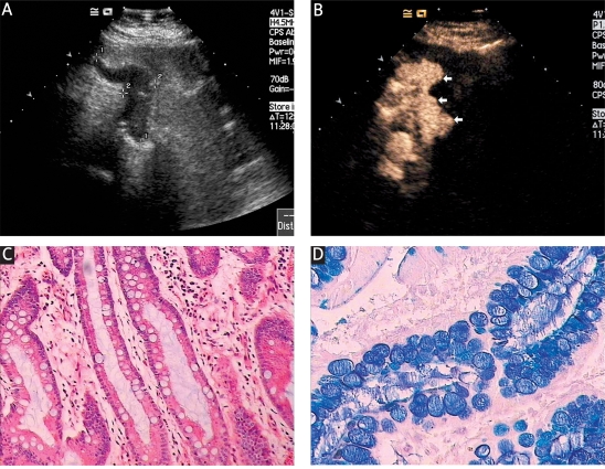
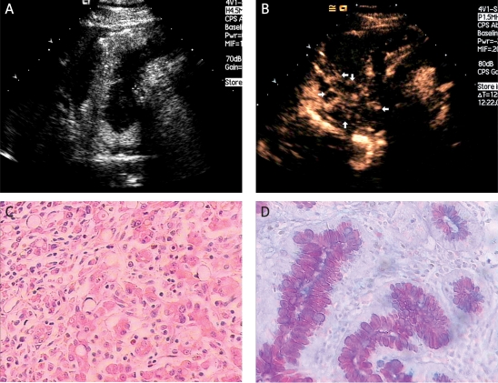
Material and methods: Fifty-eight patients with gastric cancer proved by endoscopic biopsy underwent preoperative DCUS examination in which an oral contrast agent was combined with an intravenous agent, and the findings were compared with the postoperative pathological findings using haematoxylin-eosin and Alcian Blue-Periodic Acid Schiff (AB-PAS) staining.
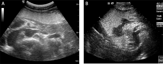
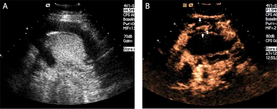
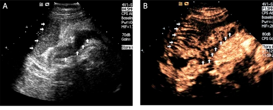
Results: Of 58 patients, 34 (59%) were the intestinal type and 24 (41%) the diffuse type on pathological examination of resected specimens. Among intestinal type patients, 30 (88%) showed homogeneous vascular enhancement and 4 (12%) heterogeneous enhancement with the "sandwich" pattern in 2 patients (50%) and "barrier" pattern in 2 patients (50%). In the diffuse type, 22 of 24 patients (92%) enhanced heterogeneously, with stippled and peripheral enhancement in 9 (41%), the "sandwich" pattern in 8 (36%) and "barrier" pattern in 5 (23%). Two of 24 patients (8%) with the diffuse type enhanced homogeneously. The proportion of heterogeneous enhancement was significantly different between the 2 subtypes of tumour (p = 0.0001). The sensitivity and specificity of heterogeneous enhancement in diagnosing the diffuse type of advanced gastric cancer were 92% and 88%, respectively. Youden's index was 0.8.
Conclusions: Double contrast-enhanced ultrasonography is a new and useful method to determine Lauren classification in patients with gastric carcinoma.
Keywords: Lauren classification; contrast; gastric carcinoma; microbubbles; ultrasonography.
Figures
References
-
- Lawrence W., Jr Gastric cancer. CA Cancer J Clin. 1986;36:216–36. - PubMed
-
- Boring CC, Squires TS, Tong T, et al. Cancer statistics. CA Cancer J Clin. 1991;41:19–36. - PubMed
-
- Parkin DM, Bray F, Ferlay J, et al. Global cancer statistics, 2002. CA Cancer J Clin. 2005;55:74–108. - PubMed
-
- Lauren P. The two histological main types of gastric carcinoma: diffuse and so-called intestinal-type carcinoma. Acta Pathol Microbiol Scand. 1965;64:31–49. - PubMed
LinkOut - more resources
Full Text Sources