

Cardiac sarcoidosis: a comprehensive review
- PMID: 22291785
- PMCID: PMC3258766
- DOI: 10.5114/aoms.2011.24118
Cardiac sarcoidosis: a comprehensive review
Abstract
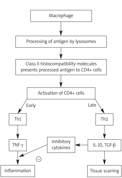
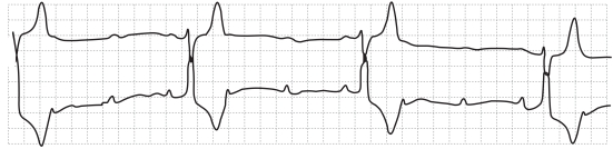
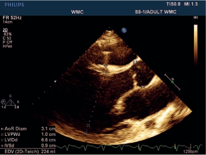
Sarcoidosis is a multisystem granulomatous disease of unknown etiology characterized by noncaseating granulomas in involved organs. Organs involved with sarcoidosis include lymph nodes, skin, lung, central nervous system, and eye. Only 40-50% of patients with cardiac sarcoidosis diagnosed at autopsy have the diagnosis made during their lifetime. Cardiac sarcoidosis can manifest itself as complete heart block, ventricular arrhythmias, congestive heart failure, pericardial effusion, pulmonary hypertension, and ventricular aneurysms. Diagnostic tests such as the electrocardiogram, two-dimensional echocardiography, cardiac magnetic resonance imaging, positron emission tomography scan, radionuclide scan, and endomyocardial biopsy can be helpful in the early detection of cardiac sarcoidosis. Considering the increased risk of sudden death, cardiac sarcoidosis is an indication for early treatment with corticosteroids or other immunosuppressive agents. Other treatments include placement of a pacemaker or implantable defibrillator to prevent sudden death. In refractory cases, cardiac transplantation should be considered.
Keywords: cardiac sarcoidosis; noncaseating granulomas; sarcoidosis.
Figures
References
-
- Benjamin AR, Marcie M, John P, et al. Racial differences in sarcoidosis incidence a 5-year study in a health maintenance organization. Am J Epidemiol. 1997;145:234–41. - PubMed
-
- Lee LS, Rose CS, Maier LA. Sarcoidosis N Engl. J Med. 1997;336:1224–34. - PubMed
-
- Bernstein M, Konzelmann FW, Sidlick DM. Boeck’s sarcoid report of a case with visceral involvement. Arch Intern Med. 1929;44:721–34.
-
- Silverman KJ, Hutchins GM, Buckley BH. Cardiac sarcoid a clinicopathologic study of 84 unselected patients with systemic sarcoidosis. Circulation. 1978;58:1204–11. - PubMed
-
- Tadamura E, Yamamuro M, Kubo S, et al. Effectiveness of delayed enhanced MRI for identification of cardiac sarcoidosis comparison with radionuclide imaging. AJR Am J Roentgenol. 2005;185:110–5. - PubMed
LinkOut - more resources
Full Text Sources