. 2012;7(1):e30517.
doi: 10.1371/journal.pone.0030517. Epub 2012 Jan 23.

Leukemia inhibitory factor in rat fetal lung development: expression and functional studies

Affiliations

Leukemia inhibitory factor in rat fetal lung development: expression and functional studies

Cristina Nogueira-Silva et al. PLoS One. 2012.

Abstract

Background: Leukemia inhibitory factor (LIF) and interleukin-6 (IL-6) are members of the family of the glycoprotein 130 (gp130)-type cytokines. These cytokines share gp130 as a common signal transducer, which explains why they show some functional redundancy. Recently, it was demonstrated that IL-6 promotes fetal lung branching. Additionally, LIF has been implicated in developmental processes of some branching organs. Thus, in this study LIF expression pattern and its effects on fetal rat lung morphogenesis were assessed.

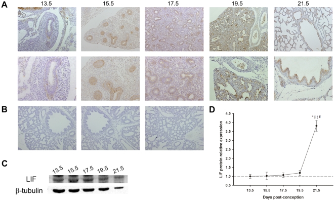
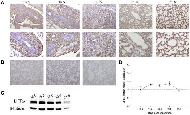
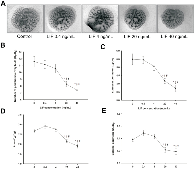
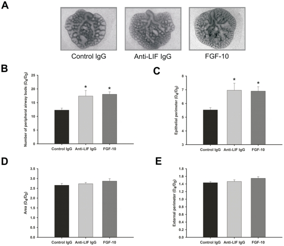
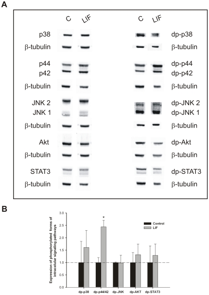
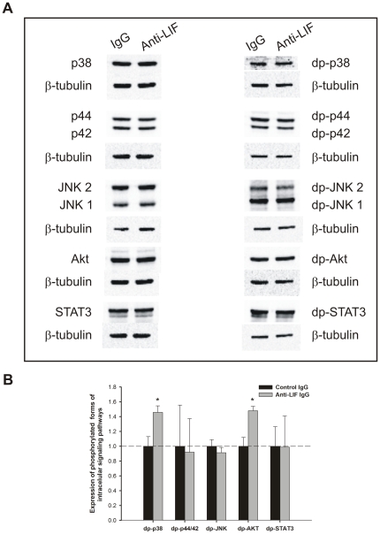
Methodology/principal findings: LIF and its subunit receptor LIFRα expression levels were evaluated by immunohistochemistry and western blot in fetal rat lungs of different gestational ages, ranging from 13.5 to 21.5 days post-conception. Throughout all gestational ages studied, LIF was constitutively expressed in pulmonary epithelium, whereas LIFRα was first mainly expressed in the mesenchyme, but after pseudoglandular stage it was also observed in epithelial cells. These results point to a LIF epithelium-mesenchyme cross-talk, which is known to be important for lung branching process. Regarding functional studies, fetal lung explants were cultured with increasing doses of LIF or LIF neutralizing antibodies during 4 days. MAPK, AKT, and STAT3 phosphorylation in the treated lung explants was analyzed. LIF supplementation significantly inhibited lung growth in spite of an increase in p44/42 phosphorylation. On the other hand, LIF inhibition significantly stimulated lung growth via p38 and Akt pathways.

Conclusions/significance: The present study describes that LIF and its subunit receptor LIFRα are constitutively expressed during fetal lung development and that they have an inhibitory physiological role on fetal lung branching.


Conflict of interest statement

Competing Interests: The authors have declared that no competing interests exist.

Figures

Figure 1
Figure 1. LIF expression pattern during fetal lung development (from 13.5 until 21.5 dpc).
(A) IHC studies revealed that LIF expression was localized to airway epithelium. Original magnification: Upper panel – ×100; Bottom panel – ×200. (B) IHC negative controls: Left – omission of the primary antibody; Center – non-immune goat IgG isotype control; Right – simultaneous omission of the primary and secondary antibodies. In all negative controls immunoreactive LIF staining was not observed. (C) Western blot analysis of LIF throughout gestation (45 kDa). Control loading was performed using β-tubulin (55 kDa). (D) Relative LIF protein levels expressed in arbitrary units normalized for β-tubulin. p<0.05: * vs. 13.5 dpc, § vs. 15.5 dpc, vs. 17.5 dpc, ¥ vs. 19.5 dpc.
Figure 2
Figure 2. LIFRα expression pattern during fetal lung development (from 13.5 until 21.5 dpc).
(A) IHC studies revealed that LIFRα was first mainly expressed in the mesenchyme, but after pseudoglandular stage it was also observed in epithelial cells throughout gestation. Original magnification: Upper panel – ×100; Bottom panel – ×200. (B) IHC negative controls: Left – omission of the primary antibody; Center – non-immune goat IgG isotype control; Right – simultaneous omission of the primary and secondary antibodies. In all negative controls immunoreactive LIFRα staining was not observed. (C) Western blot analysis of LIFRα throughout the gestation (190 kDa). Control loading was performed using β-tubulin (55 kDa). (D) Relative LIFRα protein levels expressed in arbitrary units normalized for β-tubulin. No significant difference was observed between gestational ages.
Figure 3
Figure 3. LIF supplementation studies in a fetal lung explant culture system.
(A) Representative examples of fetal lung explants treated daily with increasing concentrations of recombinant LIF, after 4 days in culture. Original magnification ×25. (B) Number of total airway buds; (C) Epithelial perimeter; (D) Area; (E) External perimeter of lung explants treated with LIF. LIF significantly inhibited lung growth. Results are expressed as ratio of day 4 (D4) and day 0 (D0) of culture (D4/D0 ratio). p<0.05: * vs. LIF at 0 ng/mL (control), § vs. LIF at 0.4 ng/mL, ¥ vs. LIF at 4 ng/mL.
Figure 4
Figure 4. LIF neutralizing studies in a fetal lung explant culture system.
(A) Representative examples of lung explants treated daily with normal IgG (control; 1 µg/mL), anti-LIF IgG (1 µg/mL) or FGF-10 (500 ng/mL), after 4 days in culture. Original magnification ×25. (B) Number of total airway buds; (C) Epithelial perimeter; (D) Area; (E) External perimeter of treated lung explants. Inhibition of LIF action significantly stimulated lung branching in a similar way than FGF-10. Results are expressed as D4/D0 ratio. p<0.05: * vs. control IgG.
Figure 5
Figure 5. Analysis of intracellular signaling pathways that mediates LIF actions on lung growth.
(A) Western blot analysis of p38, p44/42, JNK1/2, Akt and STAT3, and to diphosphorylated forms of p38 (dp-p38), p44/42 (dp-p44/42), SAPK/JNK (dp-JNK1/2), Akt (dp-Akt) and STAT3 (dp-STAT3) in control lung explants (C) and treated with LIF at 40 ng/mL (LIF). Control loading was performed using β-tubulin (55 kDa). p38 corresponds to 38 kDa. p44/42 correspond to 44 and 42 kDa, respectively. JNK1 and 2 correspond to 46 and 54 kDa, respectively. Akt corresponds to 60 kDa. STAT3 corresponds to two bands, 79 and 86 kDa. (B) Semi-quantitative analysis of expression of phosphorylated forms of these intracellular signaling pathways. Results are presented as arbitrary units normalized for β-tubulin. p<0.05: * vs. control.
Figure 6
Figure 6. Analysis of intracellular signaling pathways that mediates anti-LIF actions on lung branching.
(A) Western blot analysis of p38, p44/42, JNK1/2, Akt and STAT3, and to diphosphorylated forms of p38 (dp-p38), p44/42 (dp-p44/42), SAPK/JNK (dp-JNK1/2), Akt (dp-Akt) and STAT3 (dp-STAT3) in control IgG lung explants (IgG) and treated with anti-LIF IgG (Anti-LIF). Control loading was performed using β-tubulin (55 kDa). (B) Semi-quantitative analysis of expression of phosphorylated forms of these intracellular signaling pathways. Results are presented as arbitrary units normalized for β-tubulin. p<0.05: * vs. IgG.

References

    1. Warburton D, Bellusci S, De Langhe S, Del Moral PM, Fleury V, et al. Molecular mechanisms of early lung specification and branching morphogenesis. Pediatr Res. 2005;57:26–37. - PubMed
    1. Andrew DJ, Ewald AJ. Morphogenesis of epithelial tubes: Insights into tube formation, elongation, and elaboration. Dev Biol. 2010;341:34–55. - PMC - PubMed
    1. Nogueira-Silva C, Santos M, Baptista MJ, Moura RS, Correia-Pinto J. IL-6 is constitutively expressed during lung morphogenesis and enhances fetal lung explant branching. Pediatr Res. 2006;60:530–536. - PubMed
    1. Shimoya K, Taniguchi T, Matsuzaki N, Moriyama A, Murata Y, et al. Chorioamnionitis decreased incidence of respiratory distress syndrome by elevating fetal interleukin-6 serum concentration. Hum Reprod. 2000;15:2234–2240. - PubMed
    1. Ikegami T, Tsuda A, Karube A, Kodama H, Hirano H, et al. Effects of intrauterine IL-6 and IL-8 on the expression of surfactant apoprotein mRNAs in the fetal rat lung. Eur J Obstet Gynecol Reprod Biol. 2000;93:97–103. - PubMed

Publication types

MeSH terms

Substances