. 2012;7(1):e30528.
doi: 10.1371/journal.pone.0030528. Epub 2012 Jan 26.

Biochemical discrimination between selenium and sulfur 2: mechanistic investigation of the selenium specificity of human selenocysteine lyase

Affiliations

Biochemical discrimination between selenium and sulfur 2: mechanistic investigation of the selenium specificity of human selenocysteine lyase

Ann-Louise Johansson et al. PLoS One. 2012.

Abstract

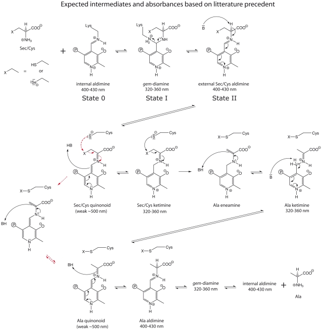
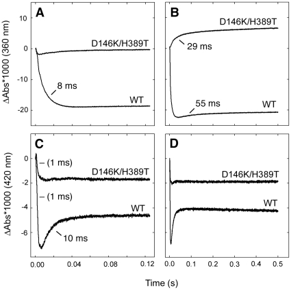
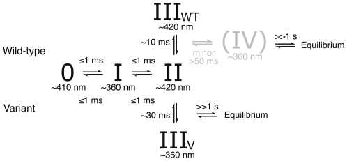
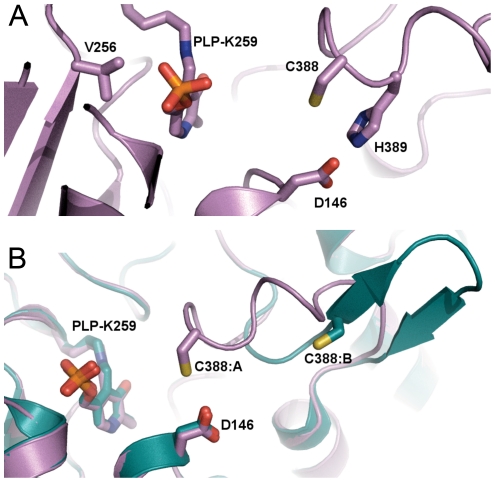
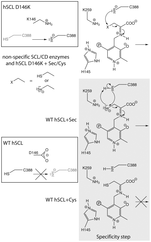
Selenium is an essential trace element incorporated into selenoproteins as selenocysteine. Selenocysteine (Sec) lyases (SCLs) and cysteine (Cys) desulfurases (CDs) catalyze the removal of selenium or sulfur from Sec or Cys, respectively, and generally accept both substrates. Intriguingly, human SCL (hSCL) is specific for Sec even though the only difference between Sec and Cys is a single chalcogen atom.The crystal structure of hSCL was recently determined and gain-of-function protein variants that also could accept Cys as substrate were identified. To obtain mechanistic insight into the chemical basis for its substrate discrimination, we here report time-resolved spectroscopic studies comparing the reactions of the Sec-specific wild-type hSCL and the gain-of-function D146K/H389T variant, when given Cys as a substrate. The data are interpreted in light of other studies of SCL/CD enzymes and offer mechanistic insight into the function of the wild-type enzyme. Based on these results and previously available data we propose a reaction mechanism whereby the Sec over Cys specificity is achieved using a combination of chemical and physico-mechanical control mechanisms.


Conflict of interest statement

Competing Interests: The authors have declared that no competing interests exist.

Figures

Figure 1
Figure 1. Consensus mechanism in SCL/CD enzymes and related systems, a variation of the mechanism, proposed in some studies is indicated by red dotted arrows , , , , , , , , , , .
Shown in the scheme are the Sec/Cys substrates, the presumed interaction with the PLP cofactor, and the active site Cys residue accepting either sulfur or selenium. The intermediate species denoted as “State 0”, “State I” and “State II” are further discussed in the text.
Figure 2
Figure 2. Spectroscopic data for the wild-type and the D146K/H389T variant proteins.
A) Spectra of as-isolated proteins taken in a scanning spectrophotometer. B–F) Diode-array stopped flow spectra 0.001, 0.5, 10, 50 and 500 sec after addition of 10 mM Cys. Reference lines are drawn at 360, 395, 410 and 420 nm.
Figure 3
Figure 3. Absorbance difference traces at 360 nm (A, B) and 420 nm (C, D) upon addition of Cys as a function of time for the wild-type and D146K/H389T variant proteins.
Panels (A, C) 0–125 ms, panels (B, D) same wavelength up to 500 ms. Note that these traces show the differences in absorbance after mixing. The absorbance at time = 0 was set to zero independently of the actual value, which means that the absolute absorbance values at different wavelengths can not be directly compared. Time constants, determed from fits with a sum of exponential functions, are also given.
Figure 4
Figure 4. Model describing the observed transitions in the reaction of WT hSCL and the D146K/H389T variant protein with excess cysteine as inferred from in the combined spectroscopic data.
Discussion of the possible nature of “State 0”, “State I” and “State II” are found in the text and also identified in Figure 1.
Figure 5
Figure 5
A) Position of residues D146K, V256S and H389T in relation to C388 and the PLP cofactor in the structure of hSCL (PDB id 3GZC). B) Superposed subunits A and B of human SCL (PDB id 3GZC) showing the structural differences in the active site segment and positioning of C388 (subunit A: pink “closed” and subunit B: cyan “open”), the location of Asp 146 is also shown.
Figure 6
Figure 6. Proposed specificity step in hSCL shown with gray background, hSCL numbering.
In non-specific enzymes the residue corresponding to 146 in hSCL is K in Group-I and H in Group-II SCL/CD proteins. The figure is drawn based on the mechanism involving elimination from the ketimine intermediate, originally proposed by Zheng et al. . However, the same reasoning is equally valid also for the alternative mechanism with elimination directly from the quinonoid intermetiate (Fig. 1, red dotted arrows).

Similar articles

Cited by

References

    1. Allmang C, Wurth L, Krol A. The selenium to selenoprotein pathway in eukaryotes: more molecular partners than anticipated. Biochim Biophys Acta. 2009;1790:1415–1423. - PubMed
    1. Yoshizawa S, Bock A. The many levels of control on bacterial selenoprotein synthesis. Biochim Biophys Acta. 2009;1790:1404–1414. - PubMed
    1. Letavayova L, Vlckova V, Brozmanova J. Selenium: from cancer prevention to DNA damage. Toxicology. 2006;227:1–14. - PubMed
    1. Schomburg L, Schweizer U, Kohrle J. Selenium and selenoproteins in mammals: extraordinary, essential, enigmatic. Cell Mol Life Sci. 2004;61:1988–1995. - PMC - PubMed
    1. Angstwurm MW, Gaertner R. Practicalities of selenium supplementation in critically ill patients. Curr Opin Clin Nutr Metab Care. 2006;9:233–238. - PubMed

Publication types

MeSH terms