

The GB virus C (GBV-C) NS3 serine protease inhibits HIV-1 replication in a CD4+ T lymphocyte cell line without decreasing HIV receptor expression
- PMID: 22292009
- PMCID: PMC3264616
- DOI: 10.1371/journal.pone.0030653
The GB virus C (GBV-C) NS3 serine protease inhibits HIV-1 replication in a CD4+ T lymphocyte cell line without decreasing HIV receptor expression
Abstract
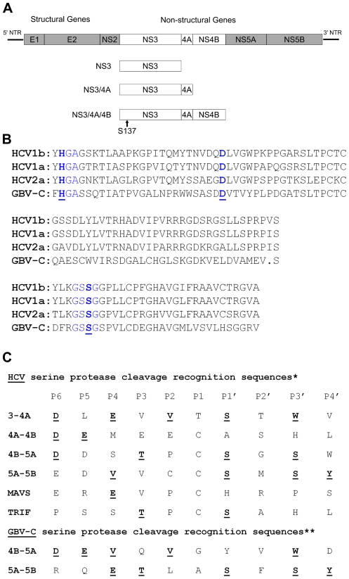
Introduction: Persistent infection with GBV-C (GB Virus C), a non-pathogenic virus related to hepatitis C virus (HCV), prolongs survival in HIV infection. Two GBV-C proteins, NS5A and E2, have been shown previously to inhibit HIV replication in vitro. We investigated whether the GBV-C NS3 serine protease affects HIV replication.
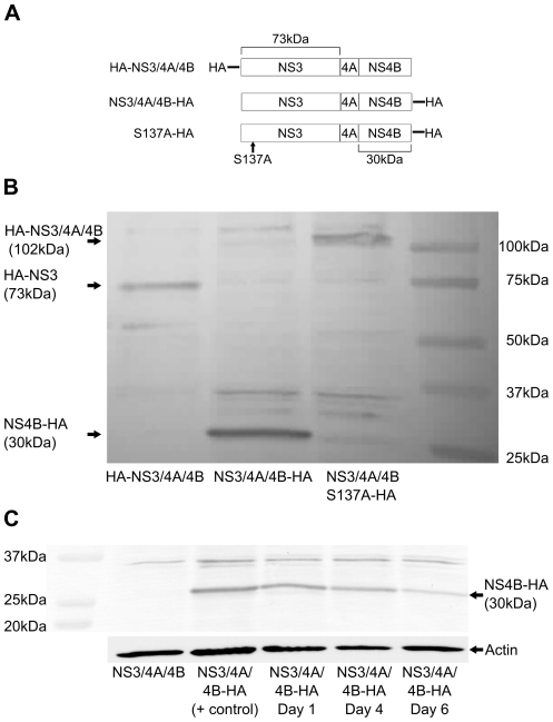
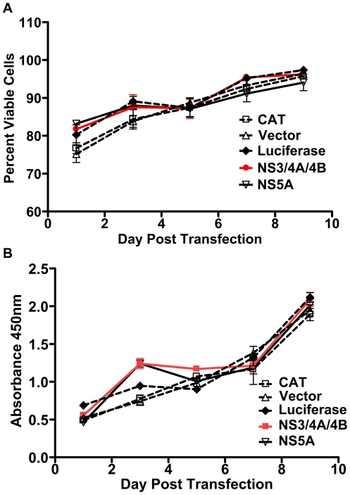
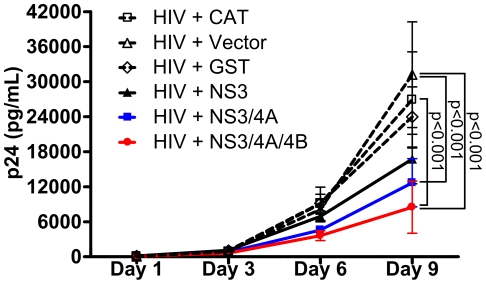
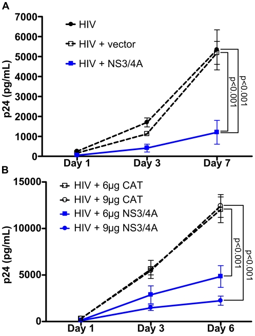
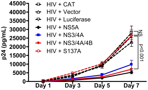
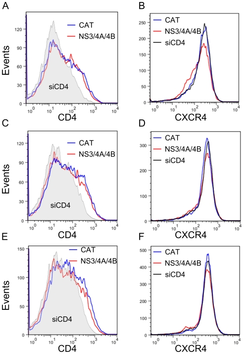
Results: GBV-C NS3 protease expressed in a human CD4+ T lymphocyte cell line significantly inhibited HIV replication. Addition of NS4A or NS4A/4B coding sequence to GBV-C NS3 increased the effect on HIV replication. Inhibition of HIV replication was dose-dependent and was not mediated by increased cell toxicity. Mutation of the NS3 catalytic serine to alanine resulted in loss of both HIV inhibition and protease activity. GBV-C NS3 expression did not measurably decrease CD4 or CXCR4 expression.
Conclusion: GBV-C NS3 serine protease significantly inhibited HIV replication without decreasing HIV receptor expression. The requirement for an intact catalytic serine at the active site indicates that inhibition was mediated by proteolytic cleavage of an unidentified target(s).
Conflict of interest statement
Figures
Similar articles
-
Virus-specific cofactor requirement and chimeric hepatitis C virus/GB virus B nonstructural protein 3.J Virol. 2000 May;74(9):4291-301. doi: 10.1128/jvi.74.9.4291-4301.2000. J Virol. 2000. PMID: 10756044 Free PMC article.
-
An 85-aa segment of the GB virus type C NS5A phosphoprotein inhibits HIV-1 replication in CD4+ Jurkat T cells.Proc Natl Acad Sci U S A. 2006 Oct 17;103(42):15570-5. doi: 10.1073/pnas.0604728103. Epub 2006 Oct 9. Proc Natl Acad Sci U S A. 2006. PMID: 17030806 Free PMC article.
-
Human pegivirus (GB virus C) NS3 protease activity inhibits induction of the type I interferon response and is not inhibited by HCV NS3 protease inhibitors.Virology. 2014 May;456-457:300-9. doi: 10.1016/j.virol.2014.03.018. Epub 2014 Apr 24. Virology. 2014. PMID: 24889249
-
GB virus type C interactions with HIV: the role of envelope glycoproteins.J Viral Hepat. 2009 Nov;16(11):757-68. doi: 10.1111/j.1365-2893.2009.01194.x. Epub 2009 Sep 15. J Viral Hepat. 2009. PMID: 19758271 Free PMC article. Review.
-
The hepatitis C virus NS3 proteinase: structure and function of a zinc-containing serine proteinase.Antivir Ther. 1998;3(Suppl 3):99-109. Antivir Ther. 1998. PMID: 10726060 Review.
Cited by
-
Clinical and molecular aspects of human pegiviruses in the interaction host and infectious agent.Virol J. 2022 Mar 9;19(1):41. doi: 10.1186/s12985-022-01769-3. Virol J. 2022. PMID: 35264187 Free PMC article. Review.
-
Detection of GB virus C genomic sequence in the cerebrospinal fluid of a HIV-infected patient in China: a case report and literature review.Epidemiol Infect. 2016 Jan;144(1):106-12. doi: 10.1017/S0950268815001326. Epub 2015 Jun 17. Epidemiol Infect. 2016. PMID: 26081197 Free PMC article. Review.
-
Human Pegivirus Type 1: A Common Human Virus That Is Beneficial in Immune-Mediated Disease?Front Immunol. 2022 May 30;13:887760. doi: 10.3389/fimmu.2022.887760. eCollection 2022. Front Immunol. 2022. PMID: 35707535 Free PMC article. Review.
-
Human pegivirus RNA is found in multiple blood mononuclear cells in vivo and serum-derived viral RNA-containing particles are infectious in vitro.J Gen Virol. 2014 Jun;95(Pt 6):1307-1319. doi: 10.1099/vir.0.063016-0. Epub 2014 Mar 25. J Gen Virol. 2014. PMID: 24668525 Free PMC article.
-
Deep sequencing identifies two genotypes and high viral genetic diversity of human pegivirus (GB virus C) in rural Ugandan patients.J Gen Virol. 2013 Dec;94(Pt 12):2670-2678. doi: 10.1099/vir.0.055509-0. Epub 2013 Sep 28. J Gen Virol. 2013. PMID: 24077364 Free PMC article.
References
-
- Xiang J, Wuenschmann S, Diekema DJ, Klinzman D, Patrick KD, et al. Effect of coinfection with GB Virus C (Hepatitis G virus) on survival among patients with HIV infection. N Eng J Med. 2001;345:707–714. - PubMed
-
- Williams CM, Klinzman D, Yamashita TE, Xiang J, Polgreen PM, et al. Persistent GB virus type C infection is associated with decreased risk of death in HIV-infection. N Eng J Med. 2003;350:981–990. - PubMed
-
- Zhang W, Chaloner K, Tillmann HL, Williams CM, Stapleton JT. Effect of early and late GB virus C viraemia on survival of HIV-infected individuals: a meta-analysis. HIV Medicine. 2006;7:173–180. - PubMed
-
- George SL, Wuenschmann S, McCoy J, Xiang J, Stapleton JT. Interactions between GB virus type C and HIV. Curr Infect Dis Reports. 2002;4:550–558. - PubMed
-
- George SL, Varmaz D. What you need to know about GB Virus C. Current Gastroenterology Reports. 2005;7:54–62. - PubMed
Publication types
MeSH terms
Substances
LinkOut - more resources
Full Text Sources
Research Materials
