. 2012;7(1):e30660.
doi: 10.1371/journal.pone.0030660. Epub 2012 Jan 23.

Hymyc1 downregulation promotes stem cell proliferation in Hydra vulgaris

Affiliations

Hymyc1 downregulation promotes stem cell proliferation in Hydra vulgaris

Alfredo Ambrosone et al. PLoS One. 2012.

Abstract

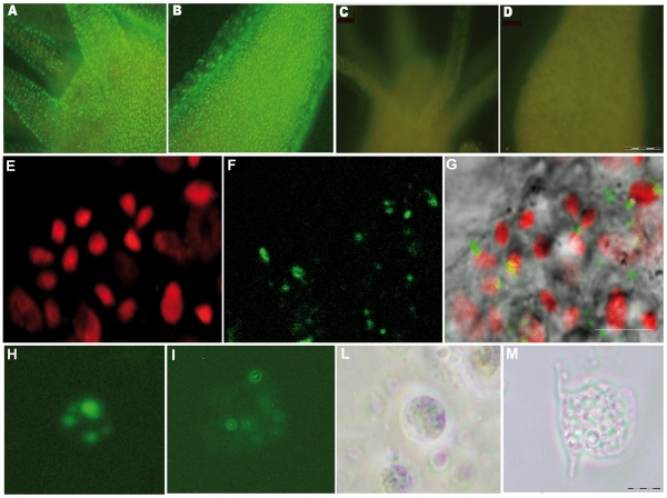
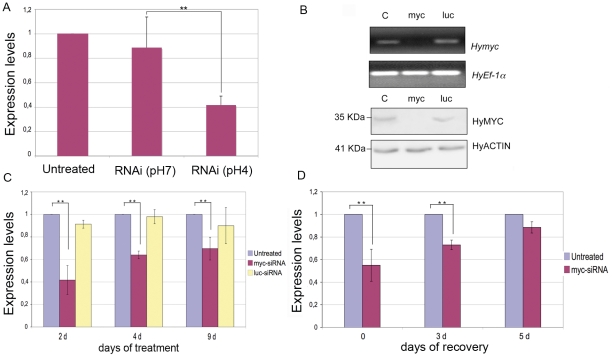
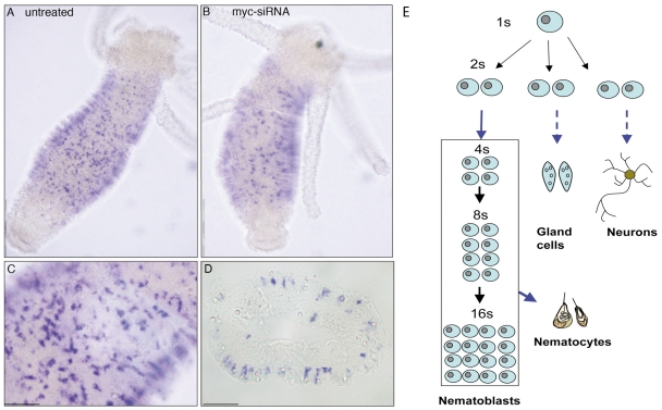
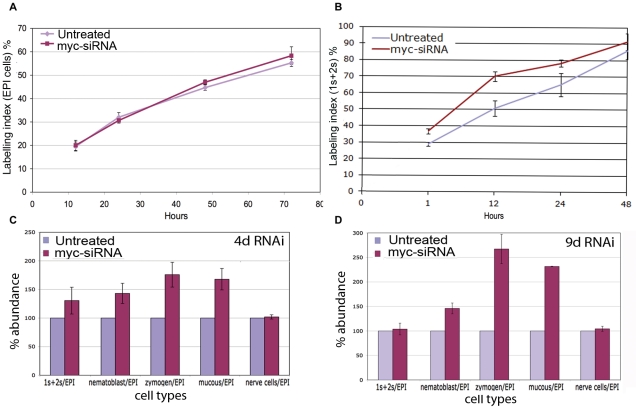
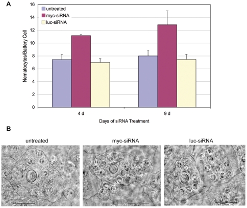
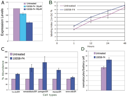
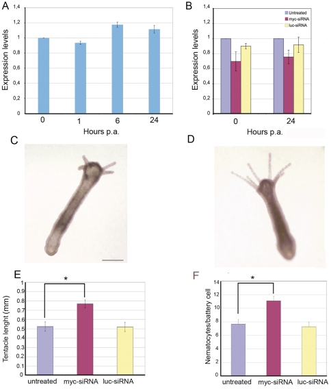
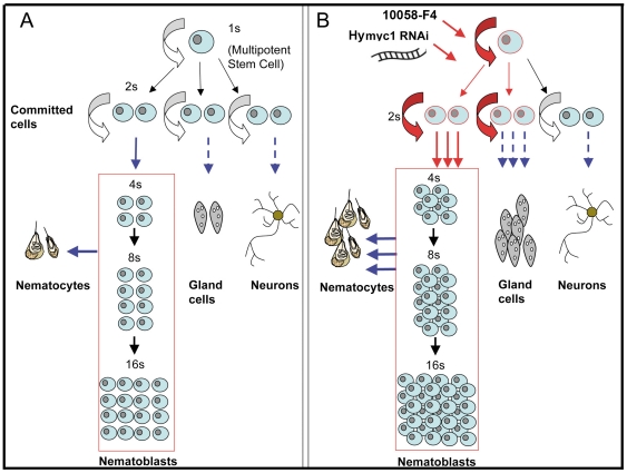
Hydra is a unique model for studying the mechanisms underlying stem cell biology. The activity of the three stem cell lineages structuring its body constantly replenishes mature cells lost due to normal tissue turnover. By a poorly understood mechanism, stem cells are maintained through self-renewal while concomitantly producing differentiated progeny. In vertebrates, one of many genes that participate in regulating stem cell homeostasis is the protooncogene c-myc, which has been recently identified also in Hydra, and found expressed in the interstitial stem cell lineage. In the present paper, by developing a novel strategy of RNA interference-mediated gene silencing (RNAi) based on an enhanced uptake of small interfering RNAi (siRNA), we provide molecular and biological evidence for an unexpected function of the Hydra myc gene (Hymyc1) in the homeostasis of the interstitial stem cell lineage. We found that Hymyc1 inhibition impairs the balance between stem cell self renewal/differentiation, as shown by the accumulation of stem cell intermediate and terminal differentiation products in genetically interfered animals. The identical phenotype induced by the 10058-F4 inhibitor, a disruptor of c-Myc/Max dimerization, demonstrates the specificity of the RNAi approach. We show the kinetic and the reversible feature of Hymyc1 RNAi, together with the effects displayed on regenerating animals. Our results show the involvement of Hymyc1 in the control of interstitial stem cell dynamics, provide new clues to decipher the molecular control of the cell and tissue plasticity in Hydra, and also provide further insights into the complex myc network in higher organisms. The ability of Hydra cells to uptake double stranded RNA and to trigger a RNAi response lays the foundations of a comprehensive analysis of the RNAi response in Hydra allowing us to track back in the evolution and the origin of this process.


Conflict of interest statement

Competing Interests: The authors have declared that no competing interests exist.

Figures

Figure 1
Figure 1. In vivo RNAi mediated by siRNA.
Fluorescence imaging of Hydra vulgaris exposed to myc-siRNA at different pHs. Living polyps were challenged with 70 nM Alexa488-end labelled myc-siRNA, in Hydra medium either at pH 4 (A, B) or at pH 7 (C, D). After 24 hr of continuous incubation with siRNA a strong punctuated fluorescence labels uniformly the whole animal, from the head (A) along the body (B) of animals treated at acidic pH, indicating the uptake of siRNA. In (C) and (D) are shown the body regions corresponding to A and B, respectively, of animals treated with the same siRNA at neutral pH. The absence of fluorescence signals indicates that the acidic pH enhances siRNA delivery in Hydra. Scale bar 200 µm. Confocal laser scanning imaging (E–G) of living Hydra treated 24 hr with Alexa488-labelled myc-siRNA revealed localization of oligonucleotides into interstitial cells. In (E), nuclear staining of interstitial cell nest with TOPO3; in (F) siRNA green fluorescence appears as faint staining or more evident granules; in (G), the overlay of bright field and fluorescence images revealed that siRNAs localize prevalently into the cytoplasm of interstitial cells. Scale bars: 10 µm. Fluorescence microscopy analysis of single cells suspensions prepared from treated dissociated animals shows intracellular localization of siRNA into interstitial (H) and epitheliomuscular (I) cells, as a punctuated pattern. The same cells were observed by phase contrast microscopy, respectively in (L) and (M). Scale bar 10 µm.
Figure 2
Figure 2. Molecular characterization of Hymyc1 RNAi.
(A) quantitative RT-PCR showing Hymyc1 downregulation enhanced by acidic condition. Animals treated by myc-siRNA at pH 4 showed 60% decrease in Hymyc1 transcription levels compared to Hydra Elongation factor HyEF1α mRNA, used as internal control (two asterisks, p<0.01 according to t-Student test). (B) Upper panel: semi-quantitative RT-PCR showing Hymyc1 downregulation induced specifically by myc-siRNA (lane myc) and not by luc-siRNA (lane luc), or in untreated animals (lane C) used as controls. Lower panel: Hymyc1 RNAi affects also MYC protein levels. Lane labels are as in the upper panel. MYC protein levels were detected using anti-HyMYC1 antibody (1∶500, kindly provided by K.Bister, University of Innsbruck) and compared to actin proteins, using an anti-actin primary antibody (1∶100, Sigma) to probe an identical blotted gel. HyMYC1 shows an apparent mol. weight of 35 kDa, as elsewhere reported . (C) Kinetics of Hymyc1 downregulation. qRT-PCR was performed on total RNA extracted from 25 animals either untreated (namely incubated at pH 4, in absence of siRNA) or incubated with the indicated siRNA for different periods. The most effective downregulation is detected at the beginning of the treatment, and it is specifically induced by myc- and not luc-siRNA duplexes (two asterisks, p<0.01 according to t-Student test). (D) Reversible effect of RNAi. Animals were treated with myc-siRNA for 4 d (time t = 0) and then cultured in physiological condition for the indicated periods of time (3 days, 5 days), when total RNAs were extracted for qRT-PCR analysis. Suspension of RNAi treatment restored in five days myc mRNA transcript levels up to physiological values. Error bars in A, C and D indicate standard deviations calculated from three independent experiments, each performed in triplicate.
Figure 3
Figure 3. Hymyc1 expression pattern is not affected by Hymyc1 downregulation.
A comparison of whole mount in situ hybridization performed on A) healthy animals and B) myc-siRNA treated animals (2 d) shows residual Hymyc1 transcripts in the RNAi targeted cells. As previously reported Hymyc1 is expressed in proliferating interstitial cells, distributed along the gastric region, shown at higher magnification in (C). A cross section of stained animals confirms this expression pattern (D). Scale bars: 1 mm in A and B; 200 µm in C and D. A scheme of the differentiation pathways in the interstitial stem cell system is shown in (E). Interstitial stem cells include multipotent stem cells (1 s) and committed stem cells (2 s) that differentiate three major cell types in Hydra (nematoblasts, secretory cells, neurons, and gametes). Nematocytes, the phylum representative cells arming the tentacles and used for pray capture, originate from nematoblasts, i.e. committed cells that after several mitotic divisions (generating nests of 4, 8 and 16 cells connected by cytoplasmic bridges) finally differentiates into several types of nematocytes (stenoteles, desmonemes, isorriza, depending on the nematocyst morphology). 2 s cells committed toward either nematocyte, nerve or secretory cells, are morphologically indistinguishable. While single interstitial stem cells (1 s) continuously self renew, among the daughter cells (2 s) 60% remain stem cells, whereas 40% become committed to different cell types.
Figure 4
Figure 4. Effect of myc RNAi on cell cycling activity and cell type distribution.
Cell cycling activity of epithelial cells (A) and 1 s+2 s stem cells (B) were obtained in control and treated animals by continuous incubation with BrdU, followed by maceration of ten animals at the indicated time points and fluorescence immunostaining. Data represent the average of three different experiments. In (C) and (D) the distribution of different cell types was assayed after 4 d and 9 d of myc RNAi, respectively. Five test animals were randomly selected from a pool of twenty five treated animals, and macerated for cell counting. The data represent the average of five independent RNAi experiments. Bars indicate standard errors. 1 s+2 s = single and pairs of interstitial cells; nematoblasts = nests of 4 s−16 s proliferating and differentiating nematoblasts; EPI = epithelial cells.
Figure 5
Figure 5. Hymyc1 RNAi induces an increase in the nematocyte content of battery cells.
(A) At time 4 d and 9 d of treatment with the indicated siRNA, animals were fixed and the number of nematocytes embedded in the battery cells, observed under the same focus plane, was scored. Data represent the average of measures from ten animals ± standard deviation. (B) Representative battery cells imaged from normal (left), myc-siRNA (middle) and luc-siRNA (right) treated animals, on whole mounts. Hand drawn black lines indicate the battery cell membranes. Hymyc1 downregulation induces nematocyte differentiation and accumulation into battery cells. Scale bar: 20 µm.
Figure 6
Figure 6. Molecular and morphological alteration induced by the c-myc inhibitor 10058-F4.
A) Living polyps were treated with the c-myc inhibitor 10058-F4 for 48 h at two different concentrations (30 µM and 90 µM) before RNA extraction for qRT-PCR analysis. Expression levels, relative to HyEF1α, indicate Hymyc1 silencing in a dose dependent manner, reaching 47% downregulation at the higher dose tested. Error bars indicate standard deviations calculated from three independent experiments, each performed in triplicate. B) Cell cycling activity of 1 s+2 s stem cells were obtained in control and treated animals (as above, 10058-F4 90 µM) by continuous incubation with BrdU, followed by maceration of ten animals at the indicated time points and fluorescence immunostaining. Labelling indexes of 4 s and gland cells are reported in Figure S6 of Supporting Information. C) Single cell suspensions were prepared from polyps treated as in A and the relative cell types counted by phase contrast microscopy. Treatment with 10058-F4 enhances stem cell proliferation and determinates the accumulation of intermediate and terminal differentiation products. D) Polyps treated with the c-myc inhibitor as in A were fixed and whole mounted for analysis of the nematocyte content into the tentacles (as described in Figure 5). The small molecule myc inhibitor induces an increase in the nematocyte content of battery cells, identical to the effect induced by myc-siRNA.
Figure 7
Figure 7. Effect of Hymyc1 RNAi on regeneration.
A) Healthy animals were bisected at time t = 0 and allowed to regenerate for the indicated period, expressed in hours. At different times post amputation (p.a.) total RNA was extracted and analysed by qRT-PCR. Not significant differences in Hymyc1 expression levels were detected during the regenerative process. B) myc-siRNA treatment (4 d) caused a reduction of myc mRNA levels. At this point (time t = 0) animals were bisected and allowed to regenerate for the indicated hours. Downregulation effects persist during regeneration. Each error bar indicates standard deviation calculated from triplicates. C–D) myc-siRNA treated animal present abnormal tentacle morphogenesis. Representative phenotypes of head regeneration at 72 hr post amputation are shown. A myc-siRNA treated animal (D) presents tentacles of increased length compared to untreated (C) or luc-siRNA treated polyps (not shown). Scale bars: 500 µm. E) The average of tentacle length, calculated on a subset of 20 animals/condition, indicates the faster tentacle development induced by myc-RNAi. Asterisk indicates p<0,05, according to t-Student test). F) The average nematocyte content of the battery cells was scored on tentacles of regenerating animals at 96 hr post amputation. myc-RNAi promotes nematocyte differentiation.
Figure 8
Figure 8. Interstitial cell differentiation pathways.
Schematic representation of the multiple differentiation pathways of the interstitial stem cells in homeostatic condition (A) and the effects on stem cell proliferation/differentiation induced by genetic or biochemical Hymyc1 downregulation (B). myc-siRNA treatment or myc biochemical inactivation induce a moderate increase in the 1 s and 2 s stem cell self-renewal and proliferation activity (red arrows). This in turn generates a higher number of both differentiating intermediates (nematoblasts) and terminal differentiated products such as nematocytes and gland cells (blue arrows). Dashed arrows indicate the absence of morphological distinct cellular intermediates. Hymyc1 downregulation does not affect the nerve differentiation pathway, consistent with the absence of Hymyc1 expression in this cell type.

Similar articles

Cited by

References

    1. Askew DS, Ashmun RA, Simmons BC, Cleveland JL. Constitutive c-myc expression in an IL-3-dependent myeloid cell line suppresses cell cycle arrest and accelerates apoptosis. Oncogene. 1991;6:1915–1922. - PubMed
    1. Bosch TC. Hydra and the evolution of stem cells. Bioessays. 2009;31:478–486. - PubMed
    1. Bode HR. The interstitial cell lineage of hydra: a stem cell system that arose early in evolution. J Cell Sci. 1996;109(Pt 6):1155–1164. - PubMed
    1. Watanabe H, Hoang VT, Mattner R, Holstein TW. Immortality and the base of multicellular life: Lessons from cnidarian stem cells. Semin Cell Dev Biol. 2009;20:1114–1125. - PubMed
    1. Campbell RD, David CN. Cell cycle kinetics and development of Hydra attenuata. II. Interstitial cells. J Cell Sci. 1974;16:349–358. - PubMed

Publication types

MeSH terms

Substances