. 2012;7(1):e30674.
doi: 10.1371/journal.pone.0030674. Epub 2012 Jan 23.

Far-infrared therapy induces the nuclear translocation of PLZF which inhibits VEGF-induced proliferation in human umbilical vein endothelial cells

Affiliations

Far-infrared therapy induces the nuclear translocation of PLZF which inhibits VEGF-induced proliferation in human umbilical vein endothelial cells

Yung-Ho Hsu et al. PLoS One. 2012.

Abstract

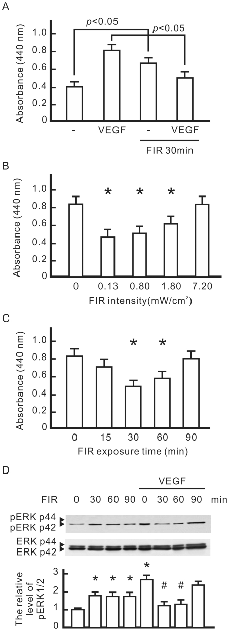
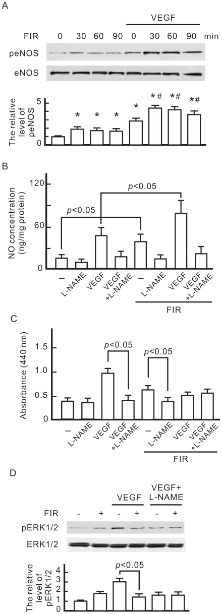
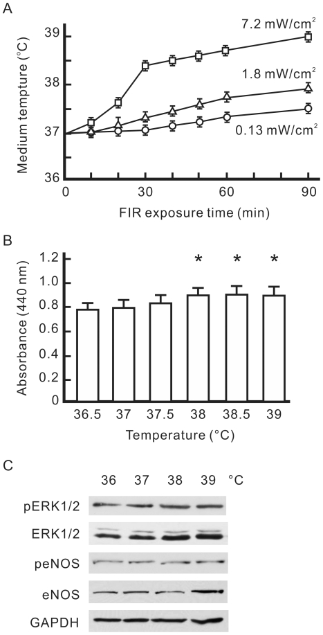
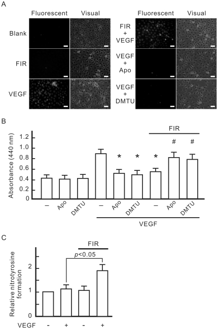
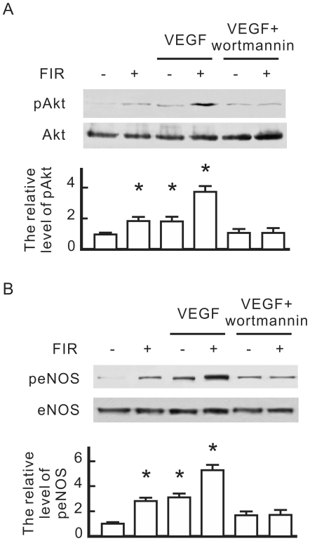
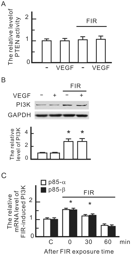
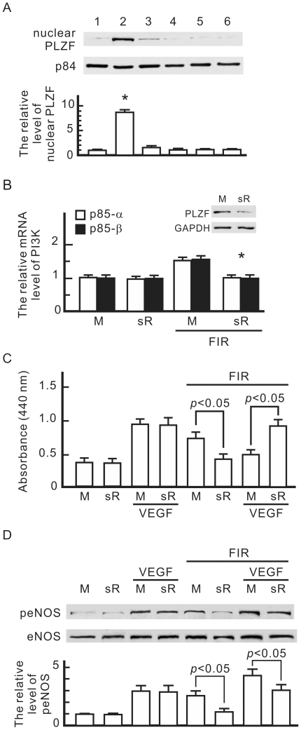
Many studies suggest that far-infrared (FIR) therapy can reduce the frequency of some vascular-related diseases. The non-thermal effect of FIR was recently found to play a role in the long-term protective effect on vascular function, but its molecular mechanism is still unknown. In the present study, we evaluated the biological effect of FIR on vascular endothelial growth factor (VEGF)-induced proliferation in human umbilical vein endothelial cells (HUVECs). We found that FIR ranging 3∼10 µm significantly inhibited VEGF-induced proliferation in HUVECs. According to intensity and time course analyses, the inhibitory effect of FIR peaked at an effective intensity of 0.13 mW/cm(2) at 30 min. On the other hand, a thermal effect did not inhibit VEGF-induced proliferation in HUVECs. FIR exposure also inhibited the VEGF-induced phosphorylation of extracellular signal-regulated kinases in HUVECs. FIR exposure further induced the phosphorylation of endothelial nitric oxide (NO) synthase (eNOS) and NO generation in VEGF-treated HUVECs. Both VEGF-induced NO and reactive oxygen species generation was involved in the inhibitory effect of FIR. Nitrotyrosine formation significantly increased in HUVECs treated with VEGF and FIR together. Inhibition of phosphoinositide 3-kinase (PI3K) by wortmannin abolished the FIR-induced phosphorylation of eNOS and Akt in HUVECs. FIR exposure upregulated the expression of PI3K p85 at the transcriptional level. We further found that FIR exposure induced the nuclear translocation of promyelocytic leukemia zinc finger protein (PLZF) in HUVECs. This induction was independent of a thermal effect. The small interfering RNA transfection of PLZF blocked FIR-increased PI3K levels and the inhibitory effect of FIR. These data suggest that FIR induces the nuclear translocation of PLZF which inhibits VEGF-induced proliferation in HUVECs.


Conflict of interest statement

Competing Interests: The authors have declared that no competing interests exist.

Figures

Figure 1
Figure 1. The influence of far infrared (FIR) exposure on VEGF-induced proliferation in HUVECs.
(A) The influence of VEGF and FIR on cell proliferation. HUVECs were pretreated with VEGF (10 ng/ml) with or without FIR exposure at 0.13 mW/cm2 for 30 min, and then cultured overnight. Cell proliferation results are presented as the absorbance of each sample at 440 nm. (B) The influence of FIR intensity on VEGF-induced cell proliferation. VEGF-pretreated cells were exposed to FIR at the indicated intensity for 30 min, and then cultured overnight. *p<0.05 vs. the control without FIR exposure. (C) The influence of FIR exposure time on VEGF-induced cell proliferation. VEGF-pretreated cells were exposed to FIR at 0.13 mW/cm2 for the indicated periods, and then cultured overnight. *p<0.05 vs. the control without FIR exposure. (D) Western blotting of VEGF-induced phosphorylation of ERK1/2. HUVECs were pretreated with or without VEGF (10 ng/ml) for 30 min, exposed to FIR at 0.13 mW/cm2 for the indicated periods, and then cultured for 8 h. ERK1/2 was detected as a loading control. By comparison with the control without FIR exposure, the relative levels of phospho-ERK1/2 were obtained and are shown as the mean±S.D. from six determinations in three cell preparations. *p<0.05 vs. the control without FIR exposure. # p<0.05 vs. the VEGF-treated group without FIR exposure.
Figure 2
Figure 2. Involvement of eNOS and NO in the biological effect of FIR in HUVECs.
(A) Western blotting of phospho-eNOS. HUVECs were pretreated with or without VEGF (10 ng/ml), exposed to FIR at 0.13 mW/cm2 for the indicated periods, and then cultured for 1 h. We detected eNOS as a loading control, and quantified phospho-eNOS expression relative to eNOS. By comparison with the control without FIR exposure, relative levels of phospho-eNOS were obtained and are shown as the mean±S.D. from six determinations in three cell preparations. *p<0.05 vs. the control with FIR exposure for 0 min. # p<0.05 vs. the VEGF-treated group with FIR exposure for 0 min. (B) Detection of FIR-induced NO. HUVECs were pretreated with VEGF and NG-nitro-L-arginine methyl ester (L-NAME) (5 mM) as indicated, exposed to FIR for 30 min, and then cultured for 1 h. The NO concentration in cultured medium was detected by a nitric oxide analyzer. (C) The influence of L-NAME on cell proliferation in HUVECs. HUVECs treated as indicated were cultured overnight. Data are shown as the mean±S.D. from six experiments. (D) Western blotting of phospho-ERK1/2. HUVECs treated as indicated were cultured for 8 h. ERK1/2 was detected as a loading control. By comparison with the control without FIR exposure, relative levels of phospho-ERK1/2 were obtained and are shown as the mean±S.D. from six determinations in three cell preparations.
Figure 3
Figure 3. Influence of a thermal effect on VEGF-induced proliferation in HUVECs.
(A) FIR exposure-increased the temperature of the culture medium. The temperature of 3 ml of culture medium in 6-cm culture dishes was detected after FIR exposure at 0.13, 1.8, or 7.2 mW/cm2 for the indicated periods. (B) The influence of thermal pretreatment on VEGF-induced cell proliferation. HUVECs were pretreated with VEGF (10 ng/ml), cultured at the indicated temperature for 30 min, and then cultured at 37°C overnight. Data are shown as the mean±S.D. from six experiments. * p<0.05 vs. the group at 37°C. (C) Western blotting of phospho-ERK1/2, ERK1/2, phospho-endothelial nitric oxide synthase (eNOS), and eNOS. HUVECs were pretreated with VEGF, cultured at the indicated temperature for 30 min, and then cultured at 37°C for 8 h. GAPDH was detected as a loading control.
Figure 4
Figure 4. Involvement of VEGF-induced ROS in the inhibitory effect of FIR on VEGF-induced proliferation in HUVECs.
(A) Detection of VEGF-induced intracellular reactive oxygen species (ROS). HUVECs were incubated for 30 min with culture medium containing DCF, and then treated with 10 ng/ml VEGF, 1 mM apocynin (Apo), 10 mM dimethylthiourea (DMTU), or FIR exposure at 0.13 mW/cm2 for 30 min as indicated. Cells were observed by fluoromicroscopy with excitation and emission wavelengths of 485 and 530 nm, respectively, and a photomicrograph is shown in the right panel of each group. Scale bar = 50 µm. (B) The influence of ROS scavengers on VEGF-induced cell proliferation. HUVECs were pretreated as indicated, and then cultured overnight. Data are shown as the mean±S.D. from six experiments. * p<0.05 vs. the group treated with VEGF alone. # p<0.05 vs. the group treated with VEGF and FIR exposure. (C) Detection of nitrotyrosine formation. HUVECs were pretreated as indicated, and then cultured for 1 h. The nitrotyrosine formation data obtained from ELISA measurements are expressed as relative quantity (means±S.D. of 9 determinations in three cell preparations).
Figure 5
Figure 5. The influence of wortmannin on FIR-induced eNOS phosphorylation in HUVECs.
HUVECs were pretreated with vascular endothelial growth factor (VEGF) (10 ng/ml), wortmannin (1 µM), or FIR exposure for 30 min as indicated, and then cultured for 1 h. (A) Western blotting of phospho-Akt. (B) Western blotting of phospho-eNOS. Akt and eNOS were detected as loading controls, and quantified phospho-eNOS and phospho-Akt expression relative to eNOS and Akt respectively. By comparison with the control without FIR exposure, relative levels of phospho-eNOS and phospho-Akt were obtained and are shown as the mean±S.D. from six determinations in three cell preparations. * p<0.05 vs. the control without FIR exposure.
Figure 6
Figure 6. The influence of FIR on the PI3K signaling pathway in HUVECs.
(A) FIR exposure-influenced phosphatase and tensin homolog (PTEN) activity. HUVECs were treated with vascular endothelial growth factor (VEGF) (10 ng/ml) or FIR exposure as indicated for 30 min. PTEN activity in each sample was detected by PTEN Malachite Green Assay Kit. The relative level of FIR-induced PTEN activity is shown as the mean±S.D. from six experiments. (B) Western blotting of PI3K p85. GAPDH was detected as a loading control. (C) FIR exposure-induced RNA levels of PI3K p85. HUVECs were pretreated with FIR exposure for 30 min, and then cultured for the indicated periods. The mRNA quantity of PI3K in each sample was detected by a qPCR with specific primers for the PI3K subunits p85α and p85β. The relative mRNA level of FIR-induced PI3K is shown as the mean±S.D. from six experiments. C, control. * p<0.05 vs. the control without FIR exposure.
Figure 7
Figure 7. Involvement of the promyelocytic leukaemia zinc finger (PLZF) in the biological effect of FIR.
(A) Western blotting of nuclear PLZF in HUVECs. HUVECs were treated as below: 1, none; 2, FIR exposure for 30 min; 3, FIR exposure for 30 min and then culture for 30 min; 4, VEGF (10 ng/ml) treatment alone for 30 min; 5, 38°C culture alone for 30 min; 6, 39°C culture alone for 30 min. Nuclear proteins were extracted and detected with a PLZF monoclonal antibody. Nuclear matrix protein p84 was detected as a loading control. By comparison with the control without FIR exposure, relative levels of nuclear PLZF were obtained and are shown as the mean±S.D. from six determinations in three cell preparations. * p<0.05 vs. the control without FIR exposure. (B) PLZF siRNA transfection-influenced RNA levels of PI3K p85. HUVECs were transfected with PLZF siRNA (sR) or mock control RNA (M). PLZF expression was monitored by a western blot analysis. Transfected cells were treated with or without FIR exposure as indicated for 30 min. The mRNA quantity of PI3K in each sample was detected by a qPCR with specific primers for PI3K subunits p85α and p85β. The relative mRNA level of FIR-induced PI3K is shown as the mean±S.D. from six experiments. * p<0.05 vs. the mock control with FIR exposure. (C) PLZF siRNA transfection-influenced cell proliferation in HUVECs. Transfected cells were pretreated with VEGF (10 ng/ml) or FIR exposure as indicated for 30 min, and then cultured overnight. Data are shown as the mean±S.D. from six experiments. (D) PLZF siRNA transfection-influenced phospho-eNOS in HUVECs. Transfected cells were treated as indicated and then cultured for 1 h. We detected eNOS as a loading control, and quantified phospho-eNOS expression relative to eNOS. By comparison with the mock control without VEGF treatment and FIR exposure, relative levels of phospho-eNOS were obtained and are shown as the mean±S.D. from six determinations in three cell preparations.

Similar articles

Cited by

References

    1. Imamura M, Biro S, Kihara T, Yoshifuku S, Takasaki K, et al. Repeated thermal therapy improves impaired vascular endothelial function in patients with coronary risk factors. J Am Coll Cardiol. 2001;38:1083–1088. - PubMed
    1. Ikeda Y, Biro S, Kamogawa Y, Yoshifuku S, Eto H, et al. Repeated sauna therapy increases arterial endothelial nitric oxide synthase expression and nitric oxide production in cardiomyopathic hamsters. Circ J. 2005;69:722–729. - PubMed
    1. Kihara T, Biro S, Ikeda Y, Fukudome T, Shinsato T, et al. Effects of repeated sauna treatment on ventricular arrhythmias in patients with chronic heart failure. Circ J. 2004;68:1146–1151. - PubMed
    1. Akasaki Y, Miyata M, Eto H, Shirasawa T, Hamada N, et al. Repeated thermal therapy up-regulates endothelial nitric oxide synthase and augments angiogenesis in a mouse model of hindlimb ischemia. Circ J. 2006;70:463–470. - PubMed
    1. Yu SY, Chiu JH, Yang SD, Hsu YC, Lui WY, et al. Biological effect of far-infrared therapy on increasing skin microcirculation in rats. Photodermatol Photoimmunol Photomed. 2006;22:78–86. - PubMed

Publication types

MeSH terms

Substances