. 2012 Apr 20;7(4):770-9.
doi: 10.1021/cb200536r. Epub 2012 Feb 15.

Chemically modified peptides targeting the PDZ domain of GIPC as a therapeutic approach for cancer

Affiliations

Chemically modified peptides targeting the PDZ domain of GIPC as a therapeutic approach for cancer

Chitta Ranjan Patra et al. ACS Chem Biol. .

Erratum in

Abstract

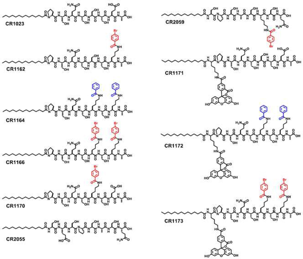
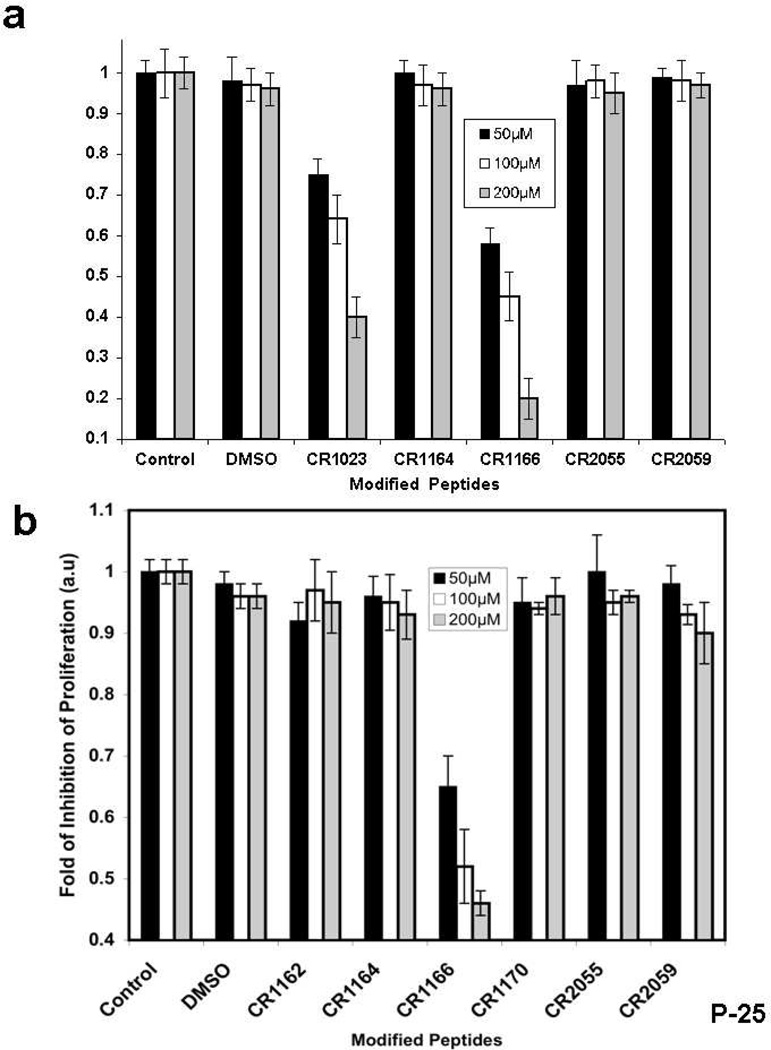
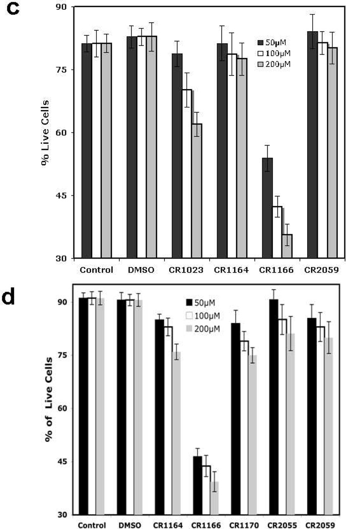
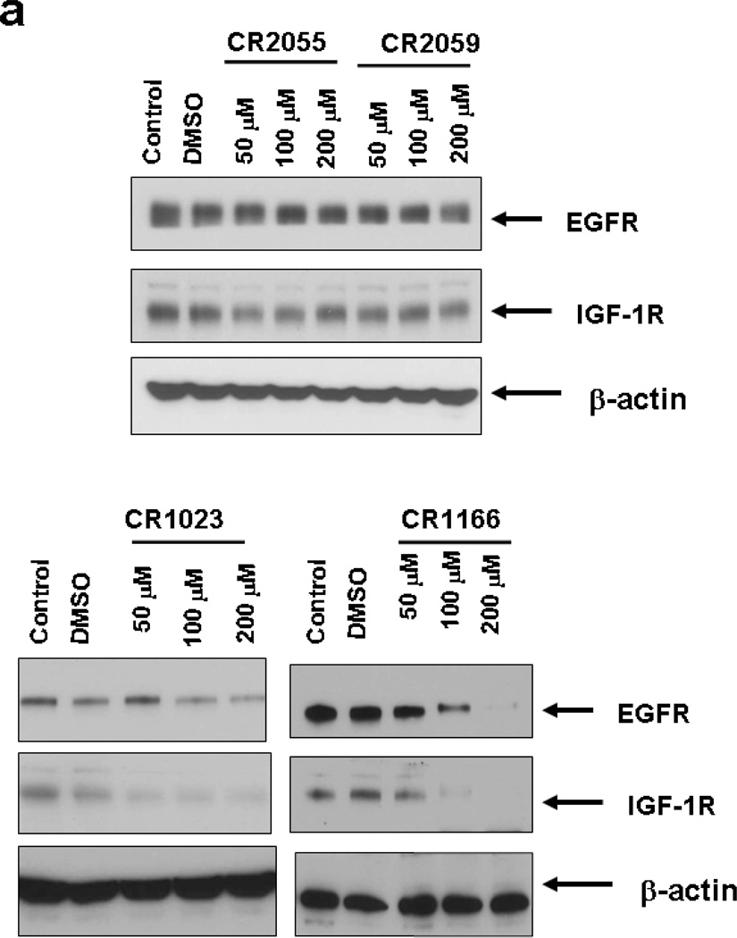
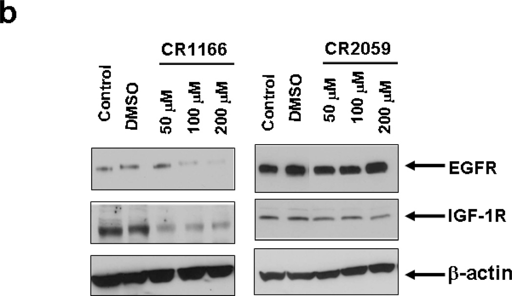
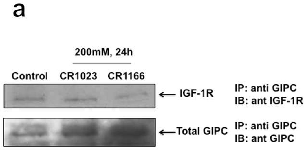
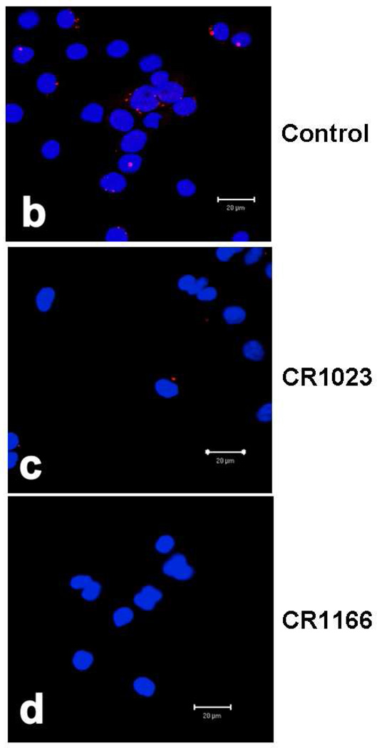
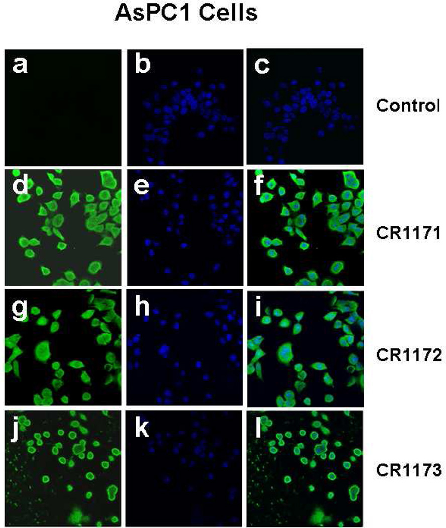
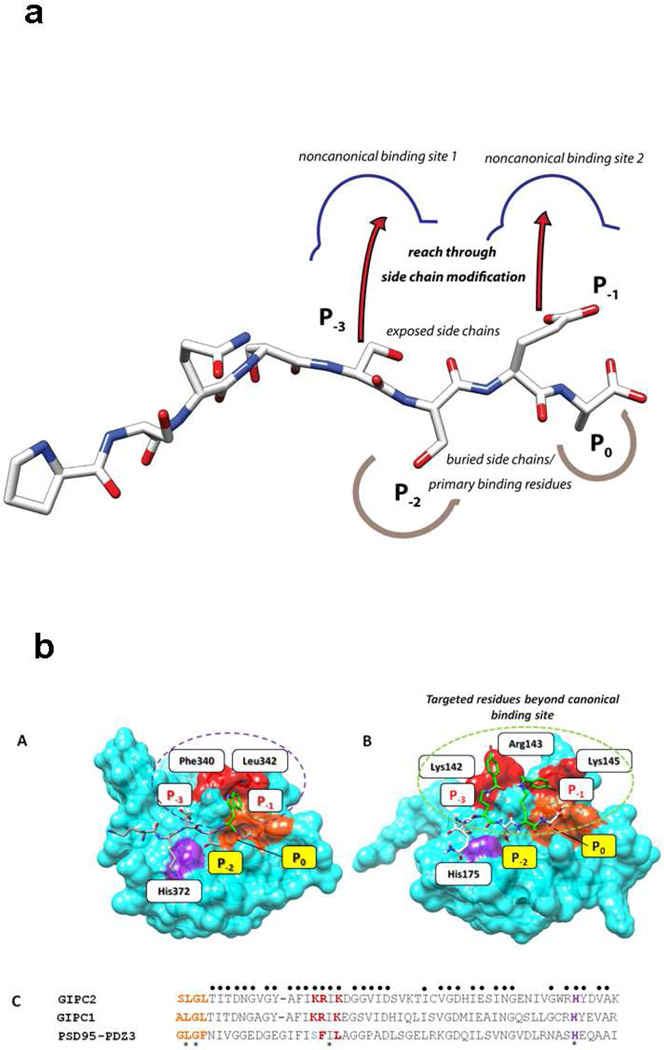
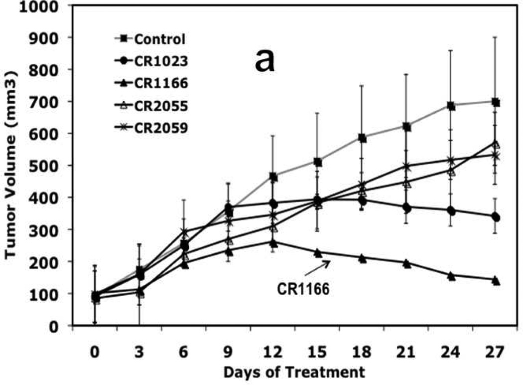
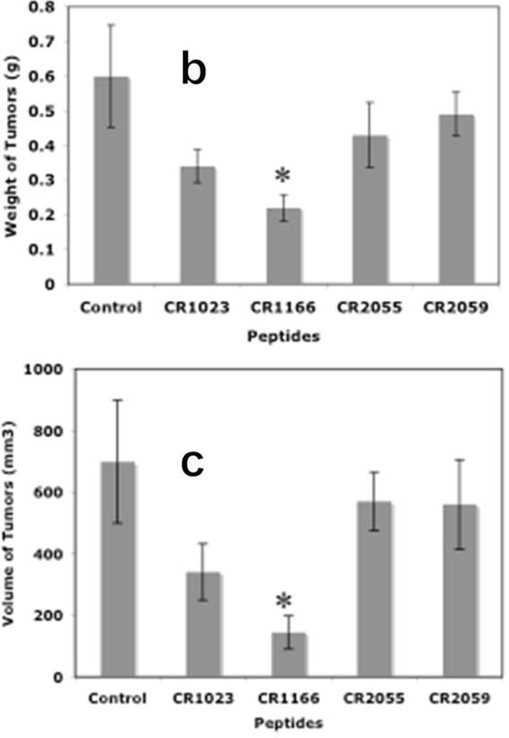
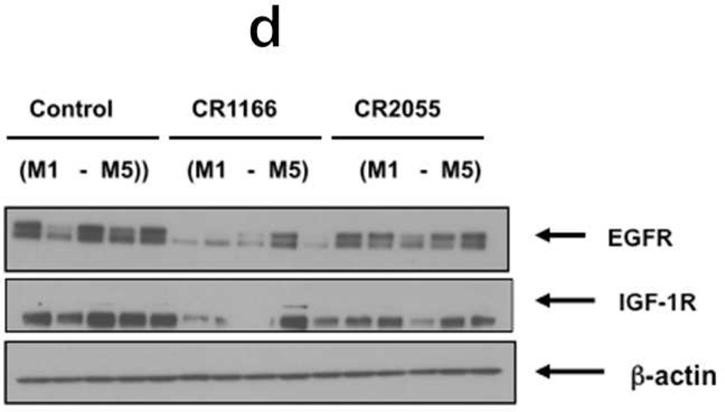
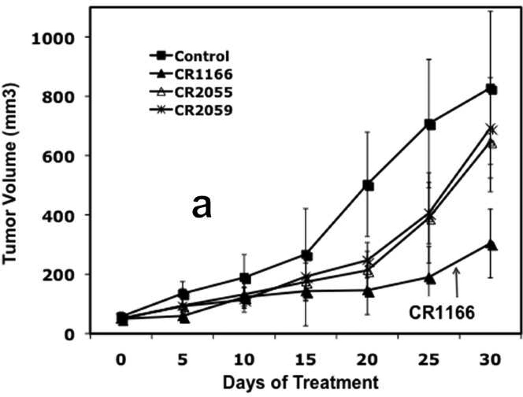
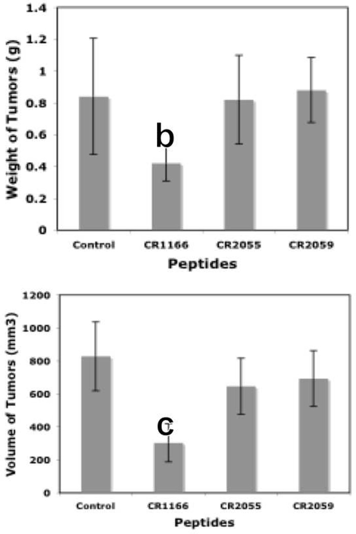
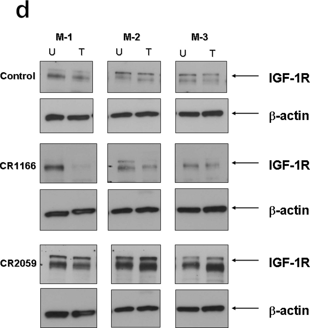
GIPC (GAIP-interacting protein, C terminus) represents a new target class for the discovery of chemotherapeutics. While many of the current generation of anticancer agents function by directly binding to intracellular kinases or cell surface receptors, the disruption of cytosolic protein-protein interactions mediated by non-enzymatic domains is an underdeveloped avenue for inhibiting cancer growth. One such example is the PDZ domain of GIPC. Previously we developed a molecular probe, the cell-permeable octapeptide CR1023 (N-myristoyl-PSQSSSEA), which diminished proliferation of pancreatic cancer cells. We have expanded upon that discovery using a chemical modification approach and here report a series of cell-permeable, side chain-modified lipopeptides that target the GIPC PDZ domain in vitro and in vivo. These peptides exhibit significant activity against pancreatic and breast cancers, both in cellular and animal models. CR1166 (N-myristoyl-PSQSK(εN-4-bromobenzoyl)SK(εN-4-bromobenzoyl)A), bearing two halogenated aromatic units on alternate side chains, was found to be the most active compound, with pronounced down-regulation of EGFR/1GF-1R expression. We hypothesize that these organic acid-modified residues extend the productive reach of the peptide beyond the canonical binding pocket, which defines the limit of accessibility for the native proteinogenic sequences that the PDZ domain has evolved to recognize. Cell permeability is achieved with N-terminal lipidation using myristate, rather than a larger CPP (cell-penetrating peptide) sequence. This, in conjunction with optimization of targeting through side chain modification, has yielded an approach that will allow the discovery and development of next-generation cellular probes for GIPC PDZ as well as for other PDZ domains.


Figures

Figure 1
Figure 1
Chemical structures of modified and unmodified peptides used in this study.
Figure 2
Figure 2. (a-d): Inhibition of proliferation and induction of apoptosis in pancreatic cancer and breast cancer: a-b
Inhibition of cell proliferation using different modified GIPC peptides in (a) AsPC1 cells and (b) MDA-MB-231-WT cells for 48 h. With a [3H]-thymidine incorporation assay, a dose-dependent significant inhibition of cell proliferation was observed in both cell lines when cells were treated with compound CR1166 compared to parent active peptide CR1023. No inhibition was observed with control peptides (CR2055, CR2059), or peptides (CR1162, CR1170) or DMSO. Inhibition of proliferation of AsPC1 cells was significantly increased with increasing concentration of CR1023 and CR1166 peptides. CR1166 is the best peptide for inhibition of cell proliferation in both cell lines compared to CR1023 and the control peptides. AsPC1 cells were incubated with different peptides for 48 hours at different three concentrations (50, 100 and 200 µM). DMSO indicates cells treated with DMSO (required for 200 µM peptides dissolved in DMSO). (c-d). Induction of apoptosis in AsPC-1 and MDA-MB-231-WT cells. Apoptosis assay of (c) AsPC-1 and (d) MDA-MB-231-WT cells treated with DMSO, and compounds CR1023, CR1164, CR1170, CR2055, and CR2059 in a dose-dependent manner (50–200µM) for 48 hours. Significant apoptosis was observed after treatment with CR1023 and CR1166 in both cell lines in a dose-dependent manner. CR1166 is most effective among all peptides. No inhibition was observed with either the control peptide CR2055 or DMSO. Apoptosis assay of AsPC-1 and MDA-MB-231-WT cells treated with DMSO and modified GIPC peptides in a dose-dependent manner (50–200µM) for 48 h of treatment using Annexin V-FITC Apoptosis Kit. Significant induction of apoptosis was observed by compound CR1166.
Figure 3
Figure 3. (a-b): Treatment of compound CR1166 down-regulates EGFR and IGF-1R expression in AsPC-1 and MDA-MB-231-WT cells
a-b. Western blot analysis of the proteins obtained from control (a) AsPC-1 cells or (b) MDA-MB-231-WT cells and cells treated with different modified Myr-GIPC peptides at different doses (50 µM – 200µM) for 48 hours. As determined by Western blotting, myristoylated GIPC peptides, most notably CR1166, regulates EGFR and IGF-1R expression in (a) AsPC-1 cells and (b) MDA-MB-231-WT cells. DMSO indicates cells treated with DMSO (required for 200 µM peptides dissolved in DMSO).
Figure 4
Figure 4. (a-d). a. Co-immunoprecipitation of IGF-IR and GIPC in AsPC1 cells
(a), immunoblot against IGF-IR after immunoprecipitation for GIPC (IP:GIPC/IB:IGF-1R). AsPC1 cells were treated with 200 µM peptide (CR1023 and CR1166) for 24 hours. This immunoblot showed that CR1166 treatment reduces the association between GIPC and IGF-IR more than CR1023 peptide. (b-d). Co-localization of GIPC and IGF-1R using CR1023 and CR1166. (b) Control, (c) treatment with CR1023, and (d) treatment with CR1166, which inhibits the association between GIPC and IGF-1R in AsPC1 cells.
Figure 5
Figure 5. (a-l). (a-l) Localization Compound CR1023, CR1164 and CR1166, in AsPC-1 cells
Confocal images of AsPC1 cells treated with FAM-conjugated peptides (CR1171, CR1172 and CR1173). (a-c) Control experiment, cells treated with DMSO, (d-f) cells treated with compound CR1171, (g-I) cells treated with compound CR1172, (j-l) cells treated with compound CR1173. Green fluorescence emissions due to presence of fluorescein in peptide were collected through a 505–550 nm band pass filter in conjunction with an argon ion laser excitation of 488 nm. The blue fluorescence emissions were collected through a 420–480 nm band pass filter in conjunction with a diode laser excitation of 405 nm for DAPI. No green fluorescence was observed in control cells treated with DMSO due to absence of fluorescein.
Figure 6
Figure 6. Design of the modified compounds based on the sequence of parent active compound, PSQSSSEA (CR1023,)
a. The P−1 and/or P−3 positions are replaced by Lys acylated with an organic acid (halogenated or non-halogenated). If only P−3 is modified, P−1 is Glu, if only P−1 position is modified, P−3 is Ser. b. Structure-based design rationale for halogenated benzoyl-modified ligands for GIPC PDZ domains, based on the complex between PDZ3 and KKETWV. Positions P0 and P−2 denote conserved primary binding determinants. (A) Key residues of PDZ3 domain binding interaction with the KKETWV are shown with conserved residues for canonical binding sites and the P−1 interaction site. (B) Expected binding mode of the pharmacophore region of CR1166 [QSK(4-bromobenzoyl)SK(4-bromobenzoyl)A] in the GIPC PDZ domain. The residues (Gly-Leu-Gly-Phe) of the carboxylate binding GLGF loop (of PDZ3) and its counterpart SLGL (in GIPC2) are colored in orange, and the His crucial for P−2 binding is shown in purple. Additional residues contacting ligands in both PDZ3 and GIPC PDZ complexes are red. The red surface represents the targeted region (P−3 and/or P−1) in PDZ proteins. In PDZ3 hydrophobic residues (Phe and Leu) line the cavity, in GIPC these are positively charged resides (Arg and Lys). In the design, either P−1 or P−3, or both, positions will be replaced by Lys acylated with halogenated benzoic acid. (C) Multiple sequence alignment of selected region of PDZ domains of GIPC2, RGS-GAIP interacting protein (GIPC1) and PSD-95-PDZ3. A sequence alignment of GIPC2, RGS-GAIP interacting protein (GIPC1) and PSD95-PDZ3 was performed with T-Coffee (www.ebi.ac.uk/t-coffee). The sequences were arranged according to structure-based alignment. The key residues identical in all three regions are marked with an asterisk, those identical to either GIPC PDZ domain regions are marked with a dot. The color scheme is as in A. The alignment scores for all three PDZ regions and for the two GIPC PDZ proteins are 81% and 98%, respectively. The amino acid sequences are as follows: GIPC2 chain A (PDB ID: 3GGE), RGS-GAIP (GIPC1) (accession AAC67550.1), and PSD95-PDZ3 (PDB ID: 1TP5). Either P−1 (Glu) or P−3 (Ser) or both positions are replaced by Lys acylated with organic acid (halogenated or non-halogenated). Structures were generated from Chem3D Pro program v11.0.1, AutoDock Tools 1.5.4 and rendered using UCSF Chimera v1.5.
Figure 7
Figure 7. (a-d), In vivo effect of intratumoral injection of peptides on tumor weight, volume, EGFR and IGF-1R expression in pancreatic tumor model
a. Effect of administration of peptides on the tumor growth over time. b-c. Tumor weight and tumor volumes decreased after peptide treatment, especially CR1166, compared to CR1023. Tumors collected after sacrificing the SCID mice and measuring with digital slide calipers using the (1/2ab2) formula. d. EGFR and IGF-1R expression is decreased in the harvest tumors after CR1166 treatment compared to tumors treated with DMSO alone.
Figure 8
Figure 8. (a-d), In vivo effect of intratumoral injection of peptides on tumor weight, volume, EGFR and IGF-1R expression in breast tumor model
a. Effect of administration of peptides on the tumor growth over time. b-c. Tumor weight and tumor volumes decreased after peptide treatment, especially CR1166, compared to control peptides. Tumors collected after sacrifice of the SCID mice and measured by digital slide calipers using the (1/2ab2) formula. d. EGFR and IGF-1R expression is decreased in the harvest tumors after CR1166 treatment (T) compared to tumors treated with DMSO alone (U) and CR2055 and CR2059.

References

    1. Anderson KE, Mack T, Silverman D. Cancer of the pancreas. 3rd ed. New York: Oxford University Press; 2006.
    1. Saif MW. Urgent Calls for Effective Earlier Disease Detection and Multi-Modal Therapeutic Strategies in Pancreatic Cancer. J. Pancreas. 2009;10:464–465.
    1. ACS Breast Cancer Facts & Figures. [accessed January14];2009–2010 http://www.cancer.org/docroot/STT/STT_0.asp.
    1. WHO, World Health Organization. Vol. 2008. World Health Organization; 2008. Agency for Research on Cancer: Fact Sheet No. 297,” July 2008.
    1. ACS, American Cancer Society, “What Are the Key Statistics fo Breast Cancer?,”. American Cancer Society. 2008 http://www.cancer.org/2008CAFFfinalsecured.pdf.

Publication types

MeSH terms

Substances