

Review
doi: 10.1172/JCI57416.
Epub 2012 Feb 1.
Epidermal barrier dysfunction and cutaneous sensitization in atopic diseases
Affiliations
- PMID: 22293182
- PMCID: PMC3266780
- DOI: 10.1172/JCI57416
Item in Clipboard
Review
Epidermal barrier dysfunction and cutaneous sensitization in atopic diseases
J Clin Invest.
2012 Feb.
Abstract
Classic atopic dermatitis is complicated by asthma, allergic rhinitis, and food allergies, cumulatively referred to as atopic diseases. Recent discoveries of mutations in the filaggrin gene as predisposing factors for atopic diseases have refocused investigators' attention on epidermal barrier dysfunction as a causative mechanism. The skin's barrier function has three elements: the stratum corneum (air-liquid barrier), tight junctions (liquid-liquid barrier), and the Langerhans cell network (immunological barrier). Clarification of the molecular events underpinning epidermal barrier function and dysfunction should lead to a better understanding of the pathophysiological mechanisms of atopic diseases.
Figures
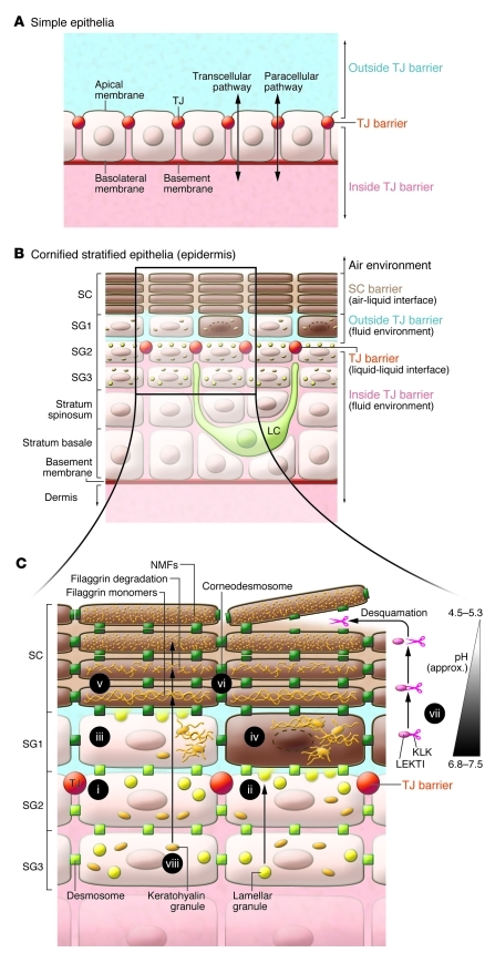
(A) In simple epithelia, TJs seal the apical end of the lateral cell membrane. The extracellular fluid is compartmentalized into two parts by TJs. (B) In the mammalian epidermis, the SC serves as an air-liquid interface barrier and protects the living layers from desiccation. TJs also seal the paracellular spaces between SG2 cells. TJs act as a liquid-liquid interface barrier in both simple and cornified stratified epithelia. LCs position their dendrites upward, ready to survey antigens upon sensing perturbation. (C) Terminal differentiation in relation to TJs. When SG3 cells differentiate into SG2 cells, they form TJs (i) and begin to secrete lamellar granules from their apical membranes (ii). SG1 cells appear to lose their TJs (iii) and then undergo final cornification (iv). Mature corneocytes are encapsulated in the cornified envelope (dark brown; v), and their intercellular spaces are filled with lipid lamellae (brown). Corneodesmosomes (green squares; vi) mediate intercorneocyte adhesion. KLKs secreted into the extracellular space are strictly limited to the extra-TJ environment. As the pH becomes acidic in the upper layers of the SC, KLKs are released from LEKTI and proteolyze corneodesmosomes, initiating desquamation (vii). Profilaggrin is a component of keratohyalin granules in the SG, is degraded into filaggrin monomers, possibly in SG1 cells, and is further degraded into NMFs in the upper SC (viii).
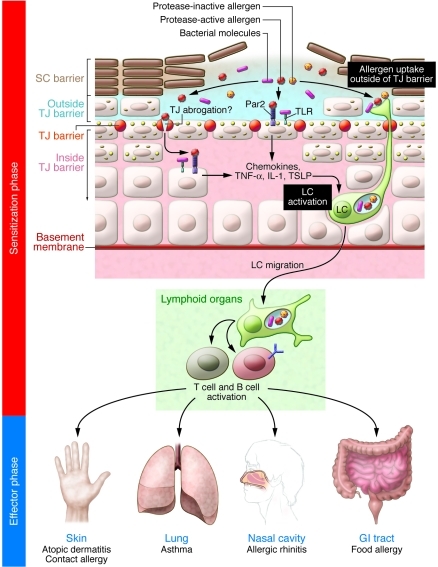
SC barrier damages induce danger signals in the epidermis. After SC barrier abrogation, protease-active allergens and uncontrolled intrinsic proteases, as well as bacterial molecules such as lipoteichoic acid of gram-positive bacteria, might agonize Par2 and TLRs on keratinocytes, respectively (7, 55, 112, 123). Keratinocytes then produce TNF-α, IL-1, and thymic stromal lymphopoietin (TSLP) (47, 55), in response to which LCs become activated (54, 56, 124). Alternatively, protease-active allergens might directly obscure the TJ barrier and then penetrate the epidermis (7), where they directly or indirectly activate LCs. Upon SC perturbation, dendrites of activated LCs penetrate the TJs to take up protease-active or -inactive antigens from the extra-TJ environment (13). Such percutaneous sensitization and chronic allergen challenge via different routes, such as the lungs, nasal cavities, and intestinal tract, are speculated to manifest as the atopic march.
References
Publication types
MeSH terms
Substances
LinkOut - more resources
Full Text Sources
Other Literature Sources
