. 2012 May;37(5):518-23.
doi: 10.1097/SHK.0b013e318249b81d.

Role of macrophages in mobilization of hematopoietic progenitor cells from bone marrow after hemorrhagic shock

Affiliations

Role of macrophages in mobilization of hematopoietic progenitor cells from bone marrow after hemorrhagic shock

Meng Xiang et al. Shock. 2012 May.

Abstract

The release of hematopoietic progenitor cells (HPCs) from bone marrow (BM) is under tight homeostatic control. Under stress conditions, HPCs migrate from BM and egress into circulation to participate in immune response, wound repair, or tissue regeneration. Hemorrhagic shock with resuscitation (HS/R), resulting from severe trauma and major surgery, promotes HPC mobilization from BM, which, in turn, affects post-HS immune responses. In this study, we investigated the mechanism of HS/R regulation of HPC mobilization from BM. Using a mouse HS/R model, we demonstrate that the endogenous alarmin molecule high-mobility group box 1 mediates HS/R-induced granulocyte colony-stimulating factor secretion from macrophages (Mϕ in a RAGE [receptor for advanced glycation end products] signaling-dependent manner. Secreted granulocyte colony-stimulating factor, in turn, induces HPC egress from BM. We also show that activation of β-adrenergic receptors on Mϕ by catecholamine mediates the HS/R-induced release of high-mobility group box 1. These data indicate that HS/R, a global ischemia-reperfusion stimulus, regulates HPC mobilization through a series of interacting pathways that include neuroendocrine and innate immune systems, in which Mϕ play a central role.


Figures
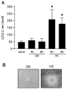
Figure 1
Figure 1. HS/R induces robust mobilization of HPC
A. HS/R increases CFU-C numbers in the blood. WT (C57BL/6) mice were subjected to HS/R (HS) or sham operation (SM). Blood samples were collected 4 and 8 h after HS/R and CFU-C assay was performed. The graph shows the mean ± SEM (n=6; *p < 0.01 compared with the groups labeled with no asterisk). B. Morphology of colonies. Colonies in HS group (4 h after HS) are much larger than that in sham mice (4 h after sham). Images are representative of six independent experiments with similar results.
Figure 2
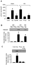
Figure 2. Catecholamine-induced HMGB1 release mediates HPC mobilization
A. Catecholamine blockade and neutralizing antibody against HMGB1 prevent HS-induced HPC mobilization. Mice were injected i.p. with neutralizing antibody against HMGB1 (Ab; 600 μg per mouse) or propranolol (Prop; 2 mg/kg) 10min before HS/R or injection of epinephrine (Ep; 2 mg/kg i.p.). Peripheral blood samples were collected 4 h after HS/R (HS) or injection of epinephrine from the right ventricle after the mice were euthanized for CFU-C assay. The graph shows the mean ± SEM (n=6; *p < 0.01 compared with the groups labeled with no asterisk). B. β-adrenergic receptor antagonist prevents HS/R-induced increase in serum HMGB1. Porpranolol (Prop; 2 mg/kg, i.p.) or epinephrine (Ep;, 2 mg/kg., i.p.) were given to WT mice 10 min before HS/R (HS) or sham operation (SM), respectively, and serum was collected after 2 h and HMGB1 detected by Western blotting. The blots are representative of 5 independent studies. The graph depicts the mean ± SEM of the fold changes in HMGB1 serum levels, as compared to sham/saline (SM/SAL) group. n= 5 mice. C. Epinephrine activation of β-adrenergic receptor induces HMGB1 release from Mφ. Mφ collected from WT mice BM were stimulated with epinephrine (Ep; 2 μg/ml) and/or propranolol (Prop; 2 μg/ml) for 2 h, HMGB1 in the supernatant was analyzed using Western blotting. Cells with no treatment are control. The blots shown are representative of three independent experiments with similar results. The graph depicts the mean ± SEM of the fold changes in HMGB1 in the supernatants as compared to the group with no treatment.
Figure 3
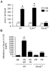
Figure 3. RAGE mediates HS/R-induced HPC mobilization
A. HMGB1 mainly acts through RAGE rather than TLR4 to induce HPC mobilization. WT (C57BL/6) mice, TLR4−/− mice and RAGE −/− mice were subjected to HS/R (HS) or sham operation (SM). Blood samples were collected at 4 h after HS/R and CFU-C assay was performed. The graph shows the mean ± SEM (n=4; *p < 0.01 compared with the groups labeled with no asterisk). B. Neutralizing antibody against HMGB1 or RAGE −/− mice attenuate HS/R-induced release of G-CSF in circulation. WT and RAGE−/− mice were injected i.p. with neutralizing antibody against HMGB1 (Ab; 600 μg per mouse) or non-specific IgG (IgG; 100 μg per mouse) 10min before HS/R. Peripheral blood samples were collected 2 h after HS/R (HS) or sham (SM) and serum G-CSF levels were determined by ELISA. Data represent mean ± SEM (n = 5 mice; *p < 0.01 compared with the groups labeled with no asterisk).
Figure 4
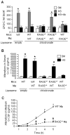
Figure 4. RAGE on Mφ mediates HMGB1-induced G-CSF release and HPC mobilization
A. Changes in HPC mobilization in chimeric mice. Mφ in WT and RAGE−/− mice were depleted using chlodronate liposome 2 days prior to HS/R, and the mice were replenished with BM Mφ isolated from WT or RAGE−/− mice and injected via tail vein (1×10⁷ cells per mouse) 15 min prior to HS/R. In some groups mice were also given with neutralizing antibody against G-CSF (Ab; 100 μg/mouse) 10 min before HS/R. Blood CFU-C was measured at 4 h after HS/R (HS) or sham operation (SM). Mice received empty liposome injection were used as a control. Data represent mean± SEM (n = 4; * p<0.01 as compared to the groups with no asterisk). B. Changes in serum G-CSF level in chimeric mice. Mφ in WT and RAGE−/− mice were depleted and repleted as described in “A”. Serum G-CSF level was measured by ELISA. Mice received empty liposome injection were used as a control. Data represent mean ± SEM (n = 4 mice; * p< 0.01 compared with the groups with no asterisk). C. In vitro Mφ release of G-CSF in response to HMGB1 stimulation. BM Mφ were isolated from either WT or RAGE−/− mice and stimulated with HMGB1 (50 μg/ml) for up to 5h. G-CSF in the cell culture medium was measured by ELISA. Data represent mean ± SEM. (n = 5 for each time point; *p<0.01 compared to WT group at that time point).

References

    1. Mendez-Ferrer S, Lucas D, Battista M, Frenette PS. Haematopoietic stem cell release is regulated by circadian oscillations. Nature. 2008;452:442–447. - PubMed
    1. Gabrilovich DI, Nagaraj S. Myeloid-derived suppressor cells as regulators of the immune system. Nat Rev Immunol. 2009;9:162–174. - PMC - PubMed
    1. Hannoush EJ, Sifri ZC, Elhassan IO, Mohr AM, Alzate WD, Offin M, Livingston DH. Impact of enhanced mobilization of bone marrow derived cells to site of injury. J Trauma. 2011;71:283–289. discussion 289–291. - PubMed
    1. Shah S, Ulm J, Sifri ZC, Mohr AM, Livingston DH. Mobilization of bone marrow cells to the site of injury is necessary for wound healing. J Trauma. 2009;67:315–321. discussion 321–312. - PubMed
    1. Livingston DH, Anjaria D, Wu J, Hauser CJ, Chang V, Deitch EA, Rameshwar P. Bone marrow failure following severe injury in humans. Ann Surg. 2003;238:748–753. - PMC - PubMed

Publication types

Substances