Review
. 2012 Mar-Apr;20(2):53-65.
doi: 10.1097/CRD.0b013e31823efde2.

Chagas heart disease: report on recent developments

Affiliations
Review

Chagas heart disease: report on recent developments

Fabiana S Machado et al. Cardiol Rev. 2012 Mar-Apr.

Abstract

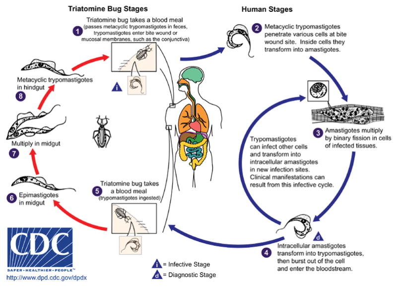
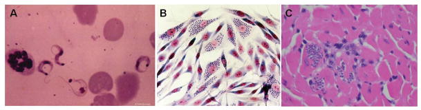
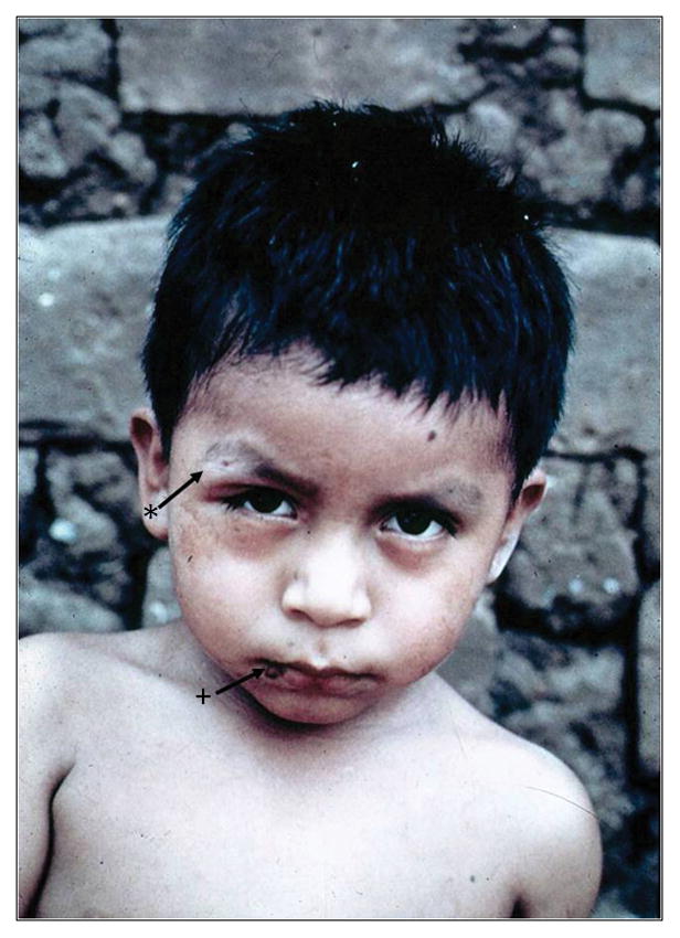
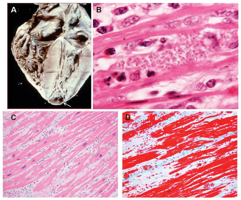
Chagas disease, caused by the parasite Trypanosoma cruzi, is an important cause of cardiac disease in endemic areas of Latin America. It is now being diagnosed in nonendemic areas because of immigration. Typical cardiac manifestations of Chagas disease include dilated cardiomyopathy, congestive heart failure, arrhythmias, cardioembolism, and stroke. Clinical and laboratory-based research to define the pathology resulting from T. cruzi infection has shed light on many of the cellular and molecular mechanisms leading to these manifestations. Antiparasitic treatment may not be appropriate for patients with advanced cardiac disease. Clinical management of Chagas heart disease is similar to that used for cardiomyopathies caused by other processes. Cardiac transplantation has been successfully performed in a small number of patients with Chagas heart disease.


Figures

Figure 1
Figure 1
Life cycle of Trypanosoma cruzi (CDC Website www.dpd.cdc.gov/dpdx/html/trypanosom/asisAmerican.htm- open access)
Figure 2
Figure 2
A Bloodform trypomastigotes B: Trypanosoma cruzi-infected myoblast culture. Note the intracellular amastigotes (arrow) C: Pseudocysts (arrow) in the myocardium of a Trypanosoma cruzi infected mouse. 2A from the collection of Herman Zaiman’s “A Presentation of Pictorial Parasites”, with permission from the American Society of Tropical Medicine and Hygiene. 2C from Tanowitz HB, Machado FS, Jelicks LA, et al: Perspectives on Trypanosoma cruzi-induced heart disease (Chagas disease). Prog Cardiovasc Dis 2009; 51: 524-39, with permission.
Figure 3
Figure 3
Child with acute Chagas disease with a chagoma (arrow with +) and Romaña’s sign (arrow with *). From the collection of Herman Zaiman’s “A Presentation of Pictorial Parasites”, with permission from the American Society of Tropical Medicine and Hygiene.
Figure 4
Figure 4
A: Heart of a patient with chronic Chagasic cardiomyopathy. There is four-chamber enlargement of the heart. Note the apical aneurysm (arrow). B: Rare pseudocyst in the myocardium of a patient with chronic Chagasic cardiomyopathy. C: H&E stained myocardium of a patient with chronic Chagasic cardiomyopathy showing bands of fibrous tissue. D: Myocardium of the same patient stained with Trichrome showing massive fibrosis. 4B from the collection of Herman Zaiman’s “A Presentation of Pictorial Parasites”, with permission from the American Society of Tropical Medicine and Hygiene.
Figure 5
Figure 5
Echocardiographic findings in Chagas heart disease. A. Transesophageal echocardiography demonstrating an apical aneurysm containing a large, round, and protruding thrombus (arrow); B–D. Transthoracic apical 4-chamber views of the heart showing dilated cardiac chambers (B), and functional mitral (C) and tricuspid (D) regurgitation (arrows). E. Parasternal short-axis view of the heart showing a large pericardial effusion (PE). F–G. Trans-mitral pulsed-Doppler (F) and lateral annulus tissue Doppler (G) demonstrating apparently normal peak early (E) and late (A) transmitral velocities, E/A ratio, and E-wave deceleration time (F) but abnormal early (E′) and late (A′) velocities (G) consistent with advanced diastolic dysfunction. LA=left atrium; LV=left ventricle; PE=pericardial effusion; RA=right atrium; RV=right ventricle. From Tanowitz HB, Machado FS, Jelicks LA, et al: Perspectives on Trypanosoma cruzi-induced heart disease (Chagas disease). Prog Cardiovasc Dis 2009; 51: 524-39, with permission.
Figure 6
Figure 6
Cardiac imaging of Trypanosoma cruzi infected mice: A: Cardiac MRI of an uninfected mouse. The right ventricle is normal in size (arrow). B: Cardiac MRI of a Trypanosoma cruzi-infected mouse 100 days post infection. Note the enlarged right ventricle (arrow). C: Cardiac microPET of an uninfected mouse showing the right ventricle (arrow). D: Cardiac microPET showing an enlarged right ventricle (arrow) in heart of a mouse 60 days post infection. From Prado et al (Ref. 139) with permission.

References

    1. Aufderheide AC, Salo W, Madden M, et al. A 9,000-year record of Chagas’ disease. Proc Natl Acad Sci U S A. 2004;101:2034–2039. - PMC - PubMed
    1. Araujo A, Jansen AM, Reinhard K, et al. Paleoparasitology of Chagas disease--a review. Mem Inst Oswaldo Cruz. 2009;104 (Suppl 1):9–16. - PubMed
    1. Biolo A, Ribeiro AL, Clausell N. Chagas cardiomyopathy--where do we stand after a hundred years? Prog Cardiovas Dis. 2010;52:300–316. - PubMed
    1. Lescure FX, Le Loup G, Freilij H, et al. Chagas disease: changes in knowledge and management. Lancet Infect Dis. 2010;10:556–570. - PubMed
    1. Rassi A, Jr, Rassi A, Marin-Neto JA. Chagas disease. Lancet. 2010;375:1388–1402. - PubMed

Publication types

MeSH terms