. 2012 Mar;5(2):207-15.
doi: 10.1038/mi.2011.67. Epub 2012 Feb 1.

c-Ets1 inhibits the interaction of NF-κB and CREB, and downregulates IL-1β-induced MUC5AC overproduction during airway inflammation

Affiliations
Free PMC article

c-Ets1 inhibits the interaction of NF-κB and CREB, and downregulates IL-1β-induced MUC5AC overproduction during airway inflammation

K S Song et al. Mucosal Immunol. 2012 Mar.
Free PMC article

Abstract

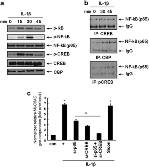
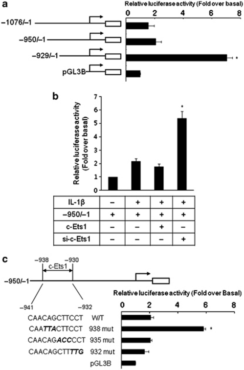
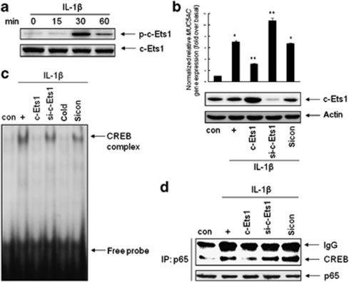
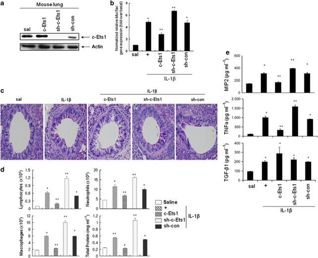
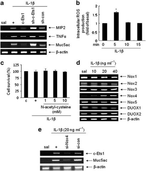
Mucin hypersecretion is frequently observed in many inflammatory diseases of the human respiratory tract. As mucin hypersecretion refers to uncontrolled mucin expression and secretion during inflammation, studies examining the negative control mechanisms of mucin hypersecretion are vital in developing novel therapeutic medications. We hypothesized that the c-Ets1 induced by interleukin (IL)-1β would decrease MUC5AC overproduction by inhibiting the interaction of NF-κB with cAMP response element-binding protein (CREB) in vivo. Stimulation with IL-1β caused the direct binding of NF-κB and CREB to the MUC5AC promoter, thus increasing MUC5AC gene expression. However, IL-1β-induced MUC5AC messenger RNA levels were surprizingly downregulated by c-Ets1 (located -938 to -930). Interestingly, c-Ets1 also suppressed IL-1β-induced MUC5AC gene expression in vitro and in vivo by disrupting the interaction of NF-κB with CREB on the MUC5AC promoter. In addition, c-Ets1 also inhibited significant morphologic changes and inflammatory cell infiltration after IL-1β exposure in mouse lungs infected with either wild-type or shRNA-c-Ets1. Moreover, reactive oxygen species produced by NOX4 increased c-Ets1 gene expression and MUC5AC gene expression in alveolar macrophages from bronchoalveolar lavage fluid. These results suggest a molecular paradigm for the establishment of a novel mechanism underlying the negative regulation of mucin overproduction, thus enhancing our understanding of airway inflammation.


Figures

Figure 1
Figure 1
Interleukin (IL)-1β induces the interaction of NF-κB with cAMP response element-binding protein (CREB) to enhance MUC5AC gene expression in NCI-H292 cells. (a) Confluent and quiescent NCI-H292 cells were treated with IL-1β (10 ng ml−1) for the indicated times, then lysates were harvested and analyzed by Western blot using several antibodies. Total NF-κB and CREB-binding protein (CBP) were used as loading controls. (b) Cells were treated for the indicated times (min) and total lysates were then immunoprecipitated with anti-CREB, CBP, or phospho-CREB antibody, and blotted with anti-p65 antibody. IgG bands were used as a loading control. IP, immunoprecipitation. (c) After cells were transfected with the small interfering RNA constructs of p65, CREB, or both, cells were treated with IL-1β for 24 h before the collection of total RNA for real-time quantitative real-time PCR. *P<0.05 compared with the control and **P<0.05 compared with IL-1β treatment only. The figures are representative of three independent experiments.
Figure 2
Figure 2
Interleukin (IL)-1β-induced MUC5AC transcription was downregulated by the cis-acting c-Ets1 regulatory motif in NCI-H292 cells. (a) The cells were transfected with several luciferase reporter constructs encoding the MUC5AC promoter and treated with IL-1β (10 ng ml−1) for 24 h. Cell lysates were analyzed with a reporter assay according to the manufacturer's instructions. *P<0.05 compared with the −950/−1 reporter construct. (b) The cells were cotransfected with pGL3B::MUC5AC −950/−1 and either the wild-type c-Ets1 expression construct or the small interfering RNA-c-Ets1 construct, and then treated with IL-1β (10 ng ml−1) for 24 h. *P<0.05 compared with the −950/−1 reporter construct. (c) Site-directed mutagenesis was carried out to generate the construct c-Ets1-binding site mutants as indicated. After treatment with IL-1β for 24 h, cell lysates were harvested. Displayed luciferase activities are shown after correction for transfection efficiency using the β-galactosidase activity of the cell lysates. Values shown are mean±s.d. of experiments performed in triplicate. *P<0.05 compared with the wild-type reporter construct.
Figure 3
Figure 3
c-Ets1 can downregulate MUC5AC gene expression by disrupting the interaction of NF-κB with cAMP response element-binding protein (CREB). (a) Confluent cells were treated with interleukin (IL)-1β (10 ng ml−1) for the indicated times, then cell lysates were analyzed by Western blot with phospho-c-Ets1 antibody. Total c-Ets1 expression was used as a loading control. (b) Cells were transiently transfected with either wild-type or the small interfering RNA (siRNA) construct of c-Ets1 or a siRNA control. Cells were serum-starved and treated with IL-1β (10 ng ml−1) for 24 h, after which cell lysates were harvested for Western blot analysis and real-time PCR. *P<0.05 compared with the control; **P<0.05 compared with IL-1β treatment. (c) Cells were transiently transfected with either the wild-type or the siRNA construct of c-Ets1 or a siRNA control. Cells were serum-starved and treated with IL-1β (10 ng ml−1) for 30 min. Nuclear protein extracts from IL-1β-treated NCI-H292 cells were subjected to electrophoretic mobility shift assay (EMSA). Nuclear proteins were incubated with [γ-32P]-labeled CRE oligonucleotides or a 50-fold excess of cold CRE probe before EMSA. The labeled nuclear proteins were separated by electrophoresis on 5% polyacrylamide gels, and the gels were dried and exposed to autoradiography at −70°C overnight. (d) Confluent and quiescent cells were transfected with either the wild-type or the siRNA construct of c-Ets1 or a siRNA control, then treated for 30 min with IL-1β. Total cell lysates were then immunoprecipitated with anti-c-Ets1 antibody and blotted with CREB antibody. IP: immunoprecipitation; IB: immunoblotting. Displayed figures are representative of three independent experiments.
Figure 4
Figure 4
c-Ets1 suppressed interleukin (IL)-1β-induced lung inflammatory responses in vivo. (a) Lenti::c-Ets1, shlenti::c-Ets1, or lenti::luciferase (multiplicity of infection 30) was administered drop by drop to the right nostril. After 3 days, lungs from the killed mice were obtained and analyzed by Western blot with c-Ets1 antibody to determine the endogenous c-Ets1 expression level (n=4). (b) Three days after infection, 50 μl of 200 ng of IL-1β or saline was instilled inside the trachea, and 24 h later, the lungs were processed for PCR. *P<0.05 compared with saline-treated mice and **P<0.05 compared with IL-1β-treated mice (n=4). Figures shown are representative of three independent experiments. (c) One week after IL-1β instillation (50 μl of 200 ng) into the lumen of the trachea of mice infected with the virus encoding wild-type c-Ets1 or shRNA-c-Ets1, periodic acid-Schiff staining was performed in the lung (n=4). (d) Mice were infected with viruses and killed 3 days post IL-1β instillation (50 μl of 200 ng), and lymphocytes, neutrophils, alveolar macrophages, and total protein levels in bronchoalveolar lavage (BAL) fluid were measured. *P<0.05 compared with saline-treated mice and **P<0.05 compared with IL-1β-treated mice (n=4). (e) The levels of MIP-2, tumor necrosis factor (TNF)-α, and transforming growth factor (TGF)-β1 proteins in BAL fluid were measured using specific enzyme-linked immunosorbent assay kits. *P<0.05 compared with saline-treated mice and **P<0.05 compared with IL-1β-treated mice (n=4).
Figure 5
Figure 5
Effect of reactive oxygen species (ROS) produced by NOX4 on c-Ets1 and MUC5AC gene expression in alveolar macrophage cells. (a) Mice were infected with viruses and killed 3 days post interleukin (IL)-1β instillation (50 μl of 200 ng), and alveolar macrophage (AM) cells from bronchoalveolar lavage fluid (n=4) were incubated. After 24 h, real-time (RT)-PCR was performed with specific primers. (b) The AM cells from saline-treated mice were pre-incubated in the presence of 50 μM 2′,7′-dichlorofluorescein diacetate for 30 min, and then exposed to IL-1β (20 ng ml−1) for the indicated periods. Cell-associated 2′,7′-dichlorofluorescein levels were analyzed by flow cytometry. *P<0.05 compared with saline-treated mice (n=4). (c) The AM cells from saline-treated mice were pre-incubated in the presence of N-acetyl-cysteine for 1 h in a dose-dependent manner, and then treated with IL-1β (20 ng ml−1) for 1 h. A cell proliferation assay was performed with CCK-8 (Dojindo, Rockville, MD) (n=4). (d) The AM cells from saline-treated mice (n=4) were exposed to IL-1β (20 ng ml−1) for the indicated times, and then were performed RT-PCR with specific primers. (e) The AM cells from saline-treated mice (n=4) were transiently transfected with either the small interfering RNA (siRNA) construct of NOX4 or a siRNA control. Cells were treated with IL-1β (20 ng ml−1) for 24 h, and RT-PCR was subsequently performed. Representative results for more than three independent experiments are shown for each group.

References

    1. Song K.S., Kim H.J., Lee J.G. Rgulator of G-protein signaling 4 suppresses LPS-induced MUC5AC overproduction in the airway. Am. J. Respir. Cell Mol. Biol. 2009;41,:40–49. - PubMed
    1. Song K.S., Choi Y.H., Kim J.M., Lee H., Lee T.J., Yoon J.H. Suppression of prostaglandin E2-induced MUC5AC overproduction by RGS4 in the airway. Am. J. Physiol. Lung Cell Mol. Physiol. 2009;296,:L684–L692. - PubMed
    1. Song K.S., Kim K., Chung K.C., Seol J.H., Yoon J.H. Interaction of SOCS3 with NonO attenuates IL-1beta-dependent MUC8 gene expression. Biochem. Biophys. Res. Commun. 2008;377,:946–951. - PubMed
    1. Fujisawa T., Velichko S., Thai P., Hung L.Y., Huang F., Wu R. Regulation of airway MUC5AC expression by IL-1beta and IL-17A; the NF-kappaB paradigm. J. Immunol. 2009;183,:6236–6243. - PMC - PubMed
    1. Song K.S., et al. Interleukin-1 beta and tumor necrosis factor-alpha induce MUC5AC overexpression through a mechanism involving ERK/p38 mitogen-activated protein kinases-MSK1-CREB activation in human airway epithelial cells. J. Biol. Chem. 2003;278,:23243–23250. - PubMed

MeSH terms