Review
. 2012 Jan 1;125(Pt 1):7-17.
doi: 10.1242/jcs.099200.

MicroRNAs and their roles in aging

Affiliations
Review

MicroRNAs and their roles in aging

Thalyana Smith-Vikos et al. J Cell Sci. .

Abstract

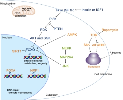
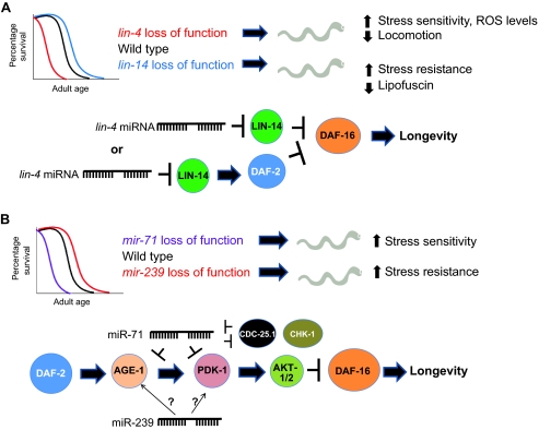
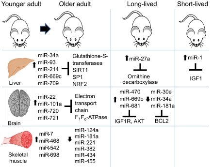
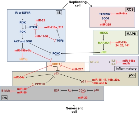
MicroRNAs (miRNAs) are a class of short non-coding RNAs that bind mRNAs through partial base-pair complementarity with their target genes, resulting in post-transcriptional repression of gene expression. The role of miRNAs in controlling aging processes has been uncovered recently with the discovery of miRNAs that regulate lifespan in the nematode Caenorhabditis elegans through insulin and insulin-like growth factor-1 signaling and DNA damage checkpoint factors. Furthermore, numerous miRNAs are differentially expressed during aging in C. elegans, but the specific functions of many of these miRNAs are still unknown. Recently, various miRNAs have been identified that are up- or down-regulated during mammalian aging by comparing their tissue-specific expression in younger and older mice. In addition, many miRNAs have been implicated in governing senescence in a variety of human cell lines, and the precise functions of some of these miRNAs in regulating cellular senescence have helped to elucidate mechanisms underlying aging. In this Commentary, we review the various regulatory roles of miRNAs during aging processes. We highlight how certain miRNAs can regulate aging on the level of organism lifespan, tissue aging or cellular senescence. Finally, we discuss future approaches that might be used to investigate the mechanisms by which miRNAs govern aging processes.


Figures

Fig. 1.
Fig. 1.
Pathways involved in aging. There are numerous cellular mechanisms that regulate aging, many of which were first shown to regulate lifespan in C. elegans (see text for details and for names of the genetic homologs in C. elegans). Regarding hormonal signaling, the IIS pathway consists of a phosphorylation cascade triggered by the insulin receptor (IR) or the IGF1 receptor (IGF1R). This cascade prevents the translocation of the FOXO transcription factor to the nucleus. When the pathway is blocked, however, FOXO functions in the nucleus to activate or repress genes important for promoting longevity. FOXO can also be regulated through phosphorylation by AMPK and JNK, and through deacetylation by SIRT1. SIRT1 activation occurs during dietary restriction, as does inhibition of TOR signaling. TOR function can also be inhibited by rapamycin; in either case, this results in an increase in autophagy, as well as a decrease in translation through activation of eIF4EBP and suppression of S6K. The FOXA and NRF transcription factors are also required to extend longevity through dietary restriction. Additionally, factors that monitor components of the mitochondrial electron transport chain and ROS generation function during aging, such as COQ7, which is required for the production of ubiquinone (coenzyme Q).
Fig. 2.
Fig. 2.
Role of miRNAs in C. elegans aging. (A) lin-4 and lin-14 regulate lifespan in C. elegans. lin-4 loss-of-function mutants display shortened lifespans and various accelerated aging phenotypes, whereas lin-14 loss-of-function mutants exhibit extended longevity [not drawn to scale; see Boehm and Slack for lifespan curves (Boehm and Slack, 2005)]. lin-4 and LIN-14 are thought to function in the same pathway as DAF-2 and DAF-16, as illustrated in the lower part of this panel. Therefore, LIN-14 function during aging might result in direct repression of DAF-16 or indirect repression of DAF-16 through DAF-2 activation, which would in either case prevent DAF-16 from activating longevity genes. (B) miR-71, 238, 239, and 246 regulate lifespan in C. elegans. mir-71 mutants (as well as mir-238 and mir-246 mutants) have a shorter lifespan than wild-type animals, whereas mir-239 mutants have a longer lifespan [not drawn to scale; see de Lencastre et al for lifespan curves (de Lencastre et al., 2010)]. Shorter-lived C. elegans also display hallmarks of accelerated aging, such as increased stress sensitivity; the opposite holds true for longer-lived animals. The targets for some of these miRNAs have been established, as illustrated in the lower part of this panel: miR-71 targets AGE-1 and PDK-1 in the IIS pathway, as well as CDC-25.1 and CDK-1, which are involved in cell cycle checkpoints for DNA damage. Additionally, miR-239 activates AGE-1 and PDK-1 through unknown mechanisms (represented with question marks on the diagram).
Fig. 3.
Fig. 3.
Role of miRNAs in aging of mice. miR-34a, miR-93, miR-214, miR-669c and miR-709 are upregulated in aged murine liver tissue. Moreover, miR-27a is increased in the liver of the long-lived Ames dwarf mouse compared with levels in wild-type mice. miR-1 is upregulated in a progeroid mouse model and targets IGF1 in the liver. In aging brain tissue, miR-22, miR-101a, miR-720 and miR-721 are upregulated. Furthermore, miR-470, miR-669b and miR-681 have increased levels in the brains of long-lived Ames dwarf mice and growth hormone receptor knockout mice, whereas miR-30e, miR-34a and miR-181a are decreased in long-lived calorie-restricted mice. Finally, miR-7, miR-468, miR-542 and miR-698 are increased in aging murine skeletal muscle, whereas miR-124a, miR-181a, miR-221, miR-382, miR-434 and miR-455 are decreased. The targets for some of these miRNAs have been identified and shown to have numerous functions during aging, such as hormonal signaling (IIS pathway), apoptosis (BCL2) and mitochondrial activity (electron transport chain components); see text for details.
Fig. 4.
Fig. 4.
miRNAs regulate factors that are important for cellular senescence. Numerous miRNAs regulate gene expression of components of aging pathways (outlined in separate boxes) on the cellular level. Stress-induced cellular senescence can be regulated by the MAPK pathway and interleukins, among other factors. Regarding MAPK, miR-15b, 24, 25 and 141 collectively target MAP2K4. The inflammatory interleukins IL6 and IL8 are secreted during stress-induced cellular senescence; miR-146a and b target and suppress these interleukins. Various miRNAs also regulate the p53 and Rb pathways. p53 activates expression of miR-34a, which targets and suppresses SIRT1 (as does miR-217), and SIRT1 normally deacetylates p53 and FOXO. Moreover, miR-29 suppresses the phosphatase PPM1D, which normally dephosphorylates p53. Furthermore, transcription of the CDK inhibitor p21 is correlated with downregulation of miR-15, miR-17, miR-19b, miR-20a and miR-106a and 106b. miR-22 targets CDK6, which then targets Rb. miR-29 and miR-30 repress the MYBL2 (B-Myb) oncogene, and their upregulation during senescence depends on Rb. The miR-17-92 cluster, as well as miR-21, miR-216a and miR-217, target PTEN, which normally blocks the phosphorylation cascade of the IIS pathway. miR-17-92 additionally target TGFβ, which also induces expression of miR-216a and miR-217. Further downstream in the IIS phosphorylation cascade, AKT induces downregulation of miR-199a-5p, which normally targets HIF1α and SIRT1. Finally, a few miRNAs regulate mitochondrial senescence-associated factors: miR-34a targets the antioxidative enzyme TXNRD2, and miR-335 targets superoxide dismutase 2 (SOD2), both of which are crucial for monitoring ROS generation.

References

    1. Bagga S., Bracht J., Hunter S., Massirer K., Holtz J., Eachus R., Pasquinelli A. E. (2005). Regulation by let-7 and lin-4 miRNAs results in target mRNA degradation. Cell 122, 553-563 - PubMed
    1. Bai X. Y., Ma Y., Ding R., Fu B., Shi S., Chen X. M. (2011). miR-335 and miR-34a promote renal senescence by suppressing mitochondrial antioxidative enzymes. J. Am. Soc. Nephrol. 22, 1252-1261 - PMC - PubMed
    1. Baker D. J., Wijshake T., Tchkonia T., LeBrasseur N. K., Childs B. G., van de Sluis B., Kirkland J. L., van Deursen J. M. (2011). Clearance of p16Ink4a-positive senescent cells delays ageing-associated disorders. Nature 479, 232-236 - PMC - PubMed
    1. Bartel D. P. (2004). MicroRNAs: Genomics, biogenesis, mechanism, and function. Cell 116, 281-297 - PubMed
    1. Bates D. J., Liang R., Li N., Wang E. (2009). The impact of noncoding RNA on the biochemical and molecular mechanisms of aging. Biochim. Biophys. Acta 1790, 970-979 - PubMed

Publication types