. 2012 Mar 6;125(9):1134-46.
doi: 10.1161/CIRCULATIONAHA.111.078212. Epub 2012 Jan 31.

Rheb is a critical regulator of autophagy during myocardial ischemia: pathophysiological implications in obesity and metabolic syndrome

Affiliations

Rheb is a critical regulator of autophagy during myocardial ischemia: pathophysiological implications in obesity and metabolic syndrome

Sebastiano Sciarretta et al. Circulation. .

Abstract

Background: Rheb is a GTP-binding protein that promotes cell survival and mediates the cellular response to energy deprivation (ED). The role of Rheb in the regulation of cell survival during ED has not been investigated in the heart.

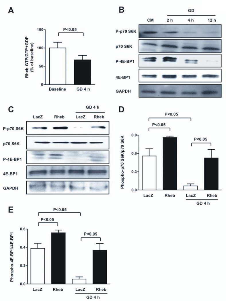
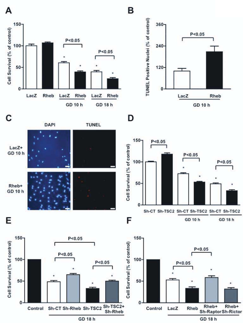
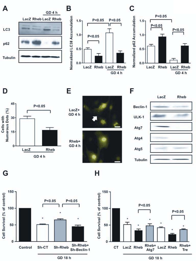
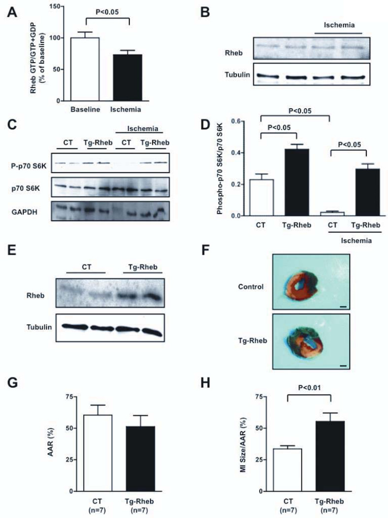
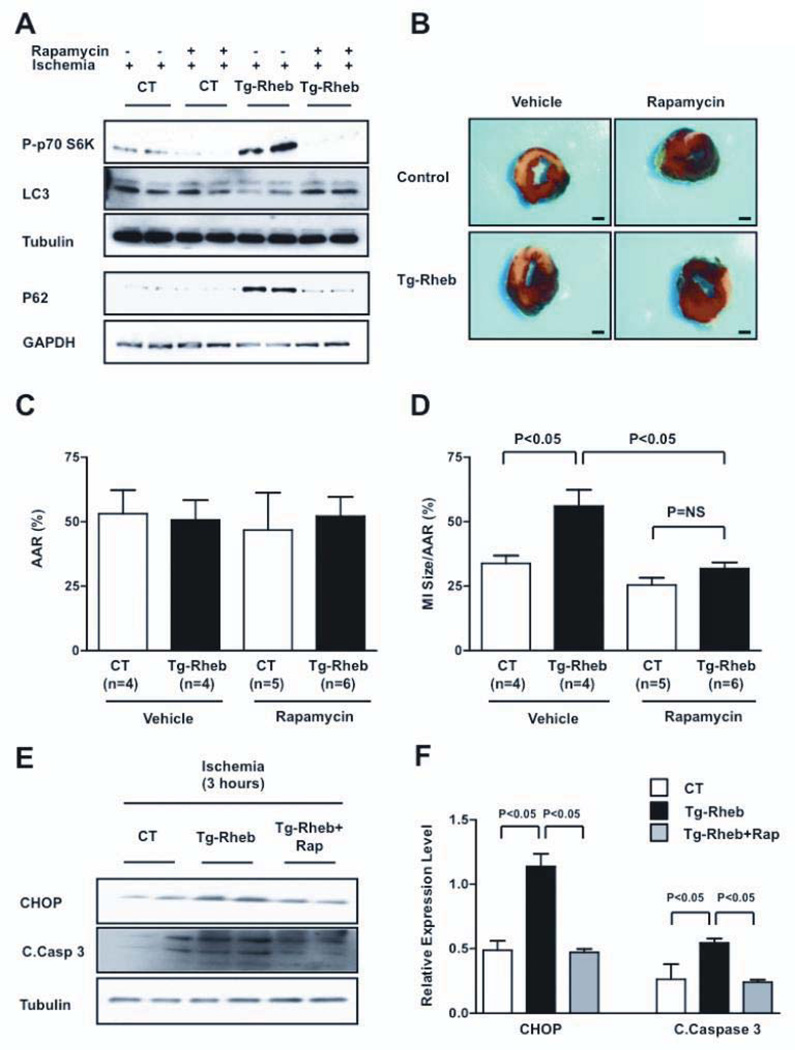
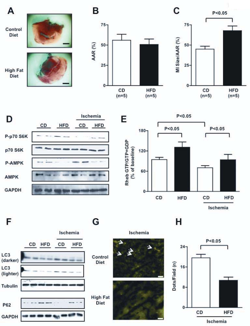
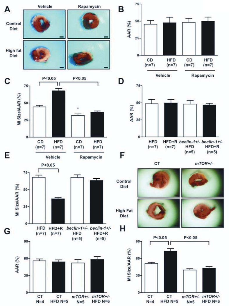
Methods and results: Rheb is inactivated during cardiomyocyte (CM) glucose deprivation (GD) in vitro, and during acute myocardial ischemia in vivo. Rheb inhibition causes mTORC1 inhibition, because forced activation of Rheb, through Rheb overexpression in vitro and through inducible cardiac-specific Rheb overexpression in vivo, restored mTORC1 activity. Restoration of mTORC1 activity reduced CM survival during GD and increased infarct size after ischemia, both of which were accompanied by inhibition of autophagy, whereas Rheb knockdown increased autophagy and CM survival. Rheb inhibits autophagy mostly through Atg7 depletion. Restoration of autophagy, through Atg7 reexpression and inhibition of mTORC1, increased cellular ATP content and reduced endoplasmic reticulum stress, thereby reducing CM death induced by Rheb activation. Mice with high-fat diet-induced obesity and metabolic syndrome (HFD mice) exhibited deregulated cardiac activation of Rheb and mTORC1, particularly during ischemia. HFD mice presented inhibition of cardiac autophagy and displayed increased ischemic injury. Pharmacological and genetic inhibition of mTORC1 restored autophagy and abrogated the increase in infarct size observed in HFD mice, but they failed to protect HFD mice in the presence of genetic disruption of autophagy.

Conclusions: Inactivation of Rheb protects CMs during ED through activation of autophagy. Rheb and mTORC1 may represent therapeutic targets to reduce myocardial damage during ischemia, particularly in obese patients.


Figures

Figure 1
Figure 1. mTORC1 is downregulated during GD through Rheb inactivation
A, Rheb activity was assessed by the ratio of Rheb-bound GTP to Rheb-bound GTP+GDP levels at baseline and during GD; N=5. B, CMs were subjected to GD for different periods of time. Phosphorylation statuses of p70S6K (Thr 389) and 4E-BP1 (Thr 37/46) were evaluated. C–E, CMs were transduced with adenovirus (Ad) harboring wild-type Rheb or LacZ. After 48 hours, phosphorylation statuses of p70S6K and 4E-BP1 were evaluated at baseline and after GD. Immunoblots and densitometric analyses are presented. N=5.
Figure 2
Figure 2. Rheb activation during GD increases CM death and apoptosis
A, Cell viability was evaluated in CMs transduced with Ad-Rheb or Ad-LacZ at baseline and during GD. N=4. B–C, Percentage of TUNEL positive cells was also evaluated after GD. N=4, bar=50 µm. D, CMs were transduced with Ad harboring short hairpin-RNA (sh)-TSC2 or with sh-scramble. After 96 hours, cell viability was evaluated at baseline and after GD. N=5. E, Cell viability was evaluated in CMs transduced with sh-scramble (sh-CT), sh-Rheb, sh-TSC2, or sh-Rheb plus sh-TSC2 at baseline and after GD. N=5. F, Cell viability was evaluated in CMs transduced with Ad-LacZ, Ad-Rheb, or Ad-Rheb plus sh-Raptor or sh-Rictor, at baseline and after GD. N=3. Cell viability was assessed by Cell Titer Blue assay. Data is presented as a percentage of the relevant control (regular medium), with the control being set at 100%. * p<0.05 with respect to relevant control (regular medium).
Figure 3
Figure 3. Rheb is a negative regulator of autophagy
A–C, CMs were transduced with Ad-LacZ or Ad-Rheb for 48 hours. LC3 isoforms and p62 accumulation were evaluated at baseline or after 4 hours of GD. A representative immunoblot is shown (A), together with densitometric analyses of LC3-II (B) and p62 (C). N=5. D–E, CMs were transduced with Ad-GFP-LC3 together with Ad-LacZ or Ad-Rheb. GFP-LC3 puncta were evaluated after 4 hours of GD (bar=10 µm). N=3. F, Expression of autophagic genes was evaluated in CMs transduced with Ad-LacZ or Ad-Rheb. G, Cell viability was evaluated in CMs transduced with sh-scramble, sh-Rheb, or with sh-Rheb plus sh-Beclin-1. N=4. Data is presented as a percentage of the relevant control (CT, baseline), with CT being set at 100%. H, Cell viability was evaluated in CMs transduced with Ad-LacZ, Ad-Rheb, and with Ad-Rheb together with Ad-Atg7 or trehalose. Sucrose treatment (100 mM) was used as control treatment for trehalose. N=3.Atg7= Ad-Atg7; Tre= trehalose. * p<0.05 with respect to cells cultured with a normal medium.
Figure 4
Figure 4. Rheb activation increases ATP depletion and ER stress during GD
A, CM ATP content at baseline and after 18 hours of GD was evaluated in CMs with Rheb overexpression or depletion. Data is presented as the fluorescence of each sample as a percentage of the control. * p<0.05 vs. control (regular medium). N=4. B–C, Unfolded protein response markers were evaluated at baseline and after GD. * p<0.05 vs. LacZ after GD. N=5. D–F, ATP content (D, N=5) and GRP78 accumulation (E–F, N=3) were evaluated in Rheb-overexpressing CMs with or without Ad-Atg7, after GD.
Figure 5
Figure 5. Rheb activation is detrimental during prolonged myocardial ischemia in vivo
A, Rheb-bound GTP levels were evaluated at baseline and after 30 minutes of ischemia. N=7. B, The amount of Rheb expression was evaluated at baseline and during ischemia. C–D, Cardiac p70S6K phosphorylation was evaluated in Tg-Rheb mice and controls, both at baseline and after 30 minutes of ischemia. N=4 for each group. E, Cardiac Rheb expression was evaluated in Tg-Rheb mice and control mice (FVB background). F–H, Tg-Rheb and control mice (Rheb+/tTA− and Rheb−/tTA+) were subjected to 3 hours of ischemia. LV myocardial sections after Alcian blue and triphenyltetrazolium chloride staining is shown (F; bar=1 mm), as well as the area at risk (AAR, G) and myocardial infarct (MI) size/AAR (H) quantification.
Figure 6
Figure 6. Rapamycin-induced autophagy limits myocardial damage in Rheb-overexpressing mice
A, Rapamycin (1 mg/kg) was administered intraperitoneally to Tg-Rheb and control mice (Rheb+/tTA−) 60 minutes before coronary ligation. p70S6K phosphorylation and LC3 expression levels were evaluated after 30 minutes of ischemia, whereas p62 expression levels were evaluated after 3 hours of ischemia. B–D, The MI /AAR ratio in Tg-Rheb and controls treated, or not treated, with rapamycin was evaluated. Bar=1 mm. E–F, CHOP expression and caspase-3 cleavage were also evaluated after 3 hours of ischemia. N=3 for each group.
Figure 7
Figure 7. HFD-induced obesity is associated with greater myocardial injury and deregulated Rheb/mTORC1 activation
A–C, MI/AAR was evaluated in CD and HFD mice after ischemia. Bar=1 mm. D, Phosphorylation statuses of p70S6K and AMPK (Thr 172) were evaluated both at baseline and after 30 minutes of ischemia (densitometric analysis is shown in Figure XIA). E, Myocardial Rheb-bound GTP content was evaluated in CD and HFD mice, both at baseline and after 30 minutes of ischemia. N=5 for each group. F, Myocardial autophagy in HFD mice was significantly inhibited compared with control mice, both at baseline and after 30 minutes (LC3-II levels) or 3 hours of ischemia (p62 levels). Representative immunoblots are presented, and densitometric analysis is reported in Supplemental Figure XIB–C. G–H, Tg-GFP-LC3 mice fed with control or high fat diet were subjected to ischemia. Representative heart sections are shown. Bar=50 µm. Arrows indicate autophagosomes (G). The number of autophagosomes per microscopic field in the two groups after 30 minutes of ischemia is reported (H). N=4 each group.
Figure 8
Figure 8. mTORC1 inhibition is protective in HFD mice through autophagy activation
A–C, Rapamycin (1 mg/kg) was administered intraperitoneally to HFD and CD mice 60 minutes before coronary ligation. Mice were subjected to 3 hours of ischemia. The MI/AAR ratio was evaluated. Bar=1 mm. * p<0.05 vs. CD. D–E, Either control or beclin-1+/− mice fed with HFD were subjected to 3 hours of ischemia. The MI/AAR ratio was evaluated. R=rapamycin. F–H, After feeding with HFD or CD, tamoxifen (30 mg/kg) was administered to α-MHC-MerCreMer-mTOR flox/+ mice (mTOR+/−) and α-MHC-MerCreMer-mTOR +/+ mice (controls) for 7 days. The mice were subjected to 3 hours of ischemia. The MI/AAR ratio was evaluated.

References

    1. Velagaleti RS, Pencina MJ, Murabito JM, Wang TJ, Parikh NI, D'Agostino RB, Levy D, Kannel WB, Vasan RS. Long-term trends in the incidence of heart failure after myocardial infarction. Circulation. 2008;118:2057–2062. - PMC - PubMed
    1. Inoki K, Ouyang H, Zhu T, Lindvall C, Wang Y, Zhang X, Yang Q, Bennett C, Harada Y, Stankunas K, Wang CY, He X, MacDougald OA, You M, Williams BO, Guan KL. TSC2 integrates Wnt and energy signals via a coordinated phosphorylation by AMPK and GSK3 to regulate cell growth. Cell. 2006;126:955–968. - PubMed
    1. Aspuria PJ, Sato T, Tamanoi F. The TSC/Rheb/TOR signaling pathway in fission yeast and mammalian cells: temperature sensitive and constitutive active mutants of TOR. Cell Cycle. 2007;6:1692–1695. - PubMed
    1. Karassek S, Berghaus C, Schwarten M, Goemans CG, Ohse N, Kock G, Jockers K, Neumann S, Gottfried S, Herrmann C, Heumann R, Stoll R. Ras homolog enriched in brain (Rheb) enhances apoptotic signaling. J Biol Chem. 2010;285:33979–33991. - PMC - PubMed
    1. Babcock JT, Quilliam LA. Rheb/mTOR Activation and Regulation in Cancer: Novel Treatment Strategies Beyond Rapamycin. Curr Drug Targets. 2011;12:1223–1231. - PubMed

Publication types

MeSH terms