. 2012 Mar 30;287(14):11351-62.
doi: 10.1074/jbc.M111.272427. Epub 2012 Jan 31.

Nurr1 protein is required for N-methyl-D-aspartic acid (NMDA) receptor-mediated neuronal survival

Affiliations

Nurr1 protein is required for N-methyl-D-aspartic acid (NMDA) receptor-mediated neuronal survival

Bruna Barneda-Zahonero et al. J Biol Chem. .

Abstract

NMDA receptor (NMDAR) stimulation promotes neuronal survival during brain development. Cerebellar granule cells (CGCs) need NMDAR stimulation to survive and develop. These neurons differentiate and mature during its migration from the external granular layer to the internal granular layer, and lack of excitatory inputs triggers their apoptotic death. It is possible to mimic this process in vitro by culturing CGCs in low KCl concentrations (5 mm) in the presence or absence of NMDA. Using this experimental approach, we have obtained whole genome expression profiles after 3 and 8 h of NMDA addition to identify genes involved in NMDA-mediated survival of CGCs. One of the identified genes was Nurr1, a member of the orphan nuclear receptor subfamily Nr4a. Our results report a direct regulation of Nurr1 by CREB after NMDAR stimulation. ChIP assay confirmed CREB binding to Nurr1 promoter, whereas CREB shRNA blocked NMDA-mediated increase in Nurr1 expression. Moreover, we show that Nurr1 is important for NMDAR survival effect. We show that Nurr1 binds to Bdnf promoter IV and that silencing Nurr1 by shRNA leads to a decrease in brain-derived neurotrophic factor (BDNF) protein levels and a reduction of NMDA neuroprotective effect. Also, we report that Nurr1 and BDNF show a similar expression pattern during postnatal cerebellar development. Thus, we conclude that Nurr1 is a downstream target of CREB and that it is responsible for the NMDA-mediated increase in BDNF, which is necessary for the NMDA-mediated prosurvival effect on neurons.


Figures

FIGURE 1.
FIGURE 1.
Gene expression profiling of NMDA treatment in CGCs. CGCs were plated in KCl 5 mm (K5) and treated or not at 2 DIV with NMDA (100 μm). After 3 and 8 h, RNA extraction was done, and samples were subjected to the Affymetrix Rat Genome 430 2.0 array. The LIMMA package was used for statistical analysis to identify up- and down-regulated genes using a multiple test (adjusted p value<0.2). A, table of the up- and down-regulated genes at 3 and 8 h classified by -fold change increase or decrease (FC = -fold change). B, pie charts representing the ontological analysis of the up-regulated genes at 8. The most enriched programs are detailed. C, heat map of the up- and down-regulated genes at 8 h. Representative genes of the synaptic transmission and antiapoptotic programs are shown.
FIGURE 2.
FIGURE 2.
Nr4a Family members are up-regulated in response to NMDA, but only Nurr1 protein levels are increased. CGCs were plated in 25 mm KCl (NT) or K5. At 2 DIV, K5 cells were treated or not with NMDA (100 μm) (N) or K25 (K). A and B, at the indicated times, RNA and cell lysates were obtained. RT-PCR (A) and Western blot (B) analyses were performed. Representative experiments are shown. Similar results were obtained in three independent experiments. C, CGCs were plated in K5. At 1 DIV, CGCs were transfected with pGL3-Basic or pGL3-Nurr1-promoter vectors, and at 2 DIV, K5 neurons were treated or not with NMDA (100 μm) or K25. Luciferase assay was performed 6 h after the treatment. Results are the mean ± S.E. of values from three independent experiments performed in duplicate. +++, p < 0.001 versus K5 pGL3-Basic; ###, p < 0.001 versus NMDA pGL3-Basic; xxx, versus K25 pGL3-Basic; ***, p < 0.001 versus K5 pGL3-Nurr1-promoter; **, p < 0.01 versus K5 pGL3-Nurr1-promoter.
FIGURE 3.
FIGURE 3.
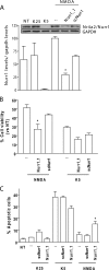
Nurr1 participates in NMDA neuroprotective effect. CGCs were plated in the presence of shRNA against Nurr1 (Nurr1_1) or a scrambled shRNA (scNurr1) as described under ”Experimental Procedures.“ At 2 DIV, cells were treated (or non-treated; NT) with NMDA (100 μm) or K25. A, upper panel, 30 h after treatment, cell lysates were obtained and subjected to Western blot with Nurr1 antibody. Lower panel, quantification of Nurr1 levels of three independent experiments are shown in the graph; *, p < 0.05 versus scNurr1. B, cell viability was assessed 7 DIV after treatment by MTT assay. Results are shown as mean ± S.E. of values from three independent experiments performed in triplicate *, p < 0.05 versus NMDA scNurr1. C, at 7 DIV, chromatin condensation was assayed by staining with Hoechst 33258. Condensed nuclei were counted and represented as percentage versus total nuclei. Results are the mean ± S.E. from three independent experiments performed in triplicate; *, p < 0.05 versus NMDA scNurr1.
FIGURE 4.
FIGURE 4.
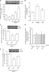
CREB regulates Nurr1 induction by NMDA. Neurons cultured in K5 were treated (or non-treated; NT) with NMDA (100 μm) or K25 at 2 DIV. A, phosphorylation of CREB (pCREB) and total CREB were determined by Western blot at the indicated times. Data are presented as mean ± S.E. of three independent experiments; ++, p < 0.01 versus K5. B, recruitment of phospho-CREB to nurr1-promoter was determined 90 min after treatment by ChIP assay (see ”Experimental Procedures“). Data represent -fold enrichment (mean ± S.E.) versus K5 condition performed in three independent experiments; **, p < 0.01 versus K5. C, D, and E, cells were plated in the presence of lentiviruses containing the indicated shRNAs and treated with NMDA at 2 DIV. Cell lysates where obtained 30 h after treatment to determine CREB (C) or Nurr1 (E) levels. A luciferase assay of Nurr1-promoter was performed as described under ”Experimental Procedures.“ D, 6 h after treatment. Data are shown as the mean ± S.E. of three or four independent experiments. **, p < 0.01, and *, p < 0.05 versus scNurr1.
FIGURE 5.
FIGURE 5.
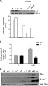
Nurr1 regulates NMDA-mediated increase in BDNF levels and has similar expression pattern to neurotrophin during postnatal cerebellum development. A, CGCs were plated in the presence or absence of the indicated shRNA and treated (or non-treated; NT) at 2 DIV with NMDA (100 μm) or K25. BDNF protein levels were assessed by Western blot 30 h after treatment. Data are from a representative experiment. Three additional independent experiments showed similar results. B, recruitment of Nurr1 to Bdnf promoters III and IV was determined 90 min after treatment by ChIP assay (see ”Experimental Procedures“). Data represent -fold enrichment (mean ± S.E.) versus K5 condition performed in three independent experiments; **, p < 0.01 versus K5. C, protein extracts from different postnatal ages of rat cerebellum were subjected to Western blot analysis to determine Nurr1 and BDNF levels. A representative Western blot is shown. Three or four animals of each age were analyzed and gave similar results.
FIGURE 6.
FIGURE 6.
Nurr1 expression is mainly restricted to nodulus lobe. Sagittal sections of cerebellum from P9, P14, and P21 rats were obtained and subjected to Nurr1 immunohistochemistry. Nurr1-increased expression during the different postnatal days was clearly evident in the nodulus lobe. Representative pictures of each age are shown (10×, 20×, 40×, and a reconstruction of whole cerebellum of each age performed with 2× photographs). The squares indicate the magnified area (nodulus), and the arrows indicate cells positive to Nurr1 staining.

Similar articles

Cited by

References

    1. Gould E., Cameron H. A., McEwen B. S. (1994) Blockade of NMDA receptors increases cell death and birth in the developing rat dentate gyrus. J. Comp. Neurol. 340, 551–565 - PubMed
    1. Ikonomidou C., Bosch F., Miksa M., Bittigau P., Vöckler J., Dikranian K., Tenkova T. I., Stefovska V., Turski L., Olney J. W. (1999) Blockade of NMDA receptors and apoptotic neurodegeneration in the developing brain. Science 283, 70–74 - PubMed
    1. Adams S. M., de Rivero Vaccari J. C., Corriveau R. A. (2004) Pronounced cell death in the absence of NMDA receptors in the developing somatosensory thalamus. J. Neurosci. 24, 9441–9450 - PMC - PubMed
    1. Balázs R., Jørgensen O. S., Hack N. (1988) N-Methyl-d-aspartate promotes the survival of cerebellar granule cells in culture. Neuroscience 27, 437–451 - PubMed
    1. Altman J. (1972) Postnatal development of the cerebellar cortex in the rat. 3: maturation of the components of the granular layer. J. Comp. Neurol. 145, 465–513 - PubMed

Publication types

MeSH terms

Substances

LinkOut - more resources